
Complete Wound Closure With ON101 Plus Standard Wound Care in Chronic Diabetic Foot Ulcers: A Multicenter U.S. Retrospective Comparative Study
Journal of Chronic Wound Care Management & Research (CWCMR) | Vol. 3, No. 1 | Published by SerenaGroup, Inc. September 1st, 2026
Bruna Nakayama1, BS; Brock A. Liden2, DPM, FAPWCA; Kylie Griffey1, BS; *Brianna Tramelli1, BS; Thomas Serena1, MD
1SerenaGroup, Inc., Norwell, MA 02061, USA; 2Wound, Ankle, Foot, and Leg, Circleville, OH, USA
* Correspondence: btramelli@serenagroups.com
ABSTRACT
Background:
Chronic diabetic foot ulcers (DFUs) remain difficult to resolve despite standard wound care. Real-world evidence describing complete wound closure and the temporal pattern of wound resolution may provide clinically relevant information regarding treatment performance outside randomized clinical trials. This study compared wound closure outcomes among patients with DFUs treated with ON101 plus standard wound care with an external historical standard-of-care (SOC) cohort.
Methods:
This multicenter retrospective observational study included 20 DFUs from 19 US patients treated with ON101 plus standard wound care and 40 de-identified DFUs from a nonconcurrent historical SOC cohort. The primary endpoint was complete wound closure by Week 12.
Secondary and exploratory outcomes included serial closure rates, Week 4 percent area reduction (PAR), achievement of ≥50% PAR at Week 4, and time to complete closure. Week 4 PAR was analyzed among evaluable wounds, with a missing-as-nonresponder sensitivity analysis. Between-cohort comparisons were performed for continuous outcomes and categorical outcomes. Time to complete closure through Week 12 was evaluated using time-to-event methods. In the ON101 cohort, the positive predictive value (PPV) of achieving ≥50% PAR at Week 4 for subsequent complete wound closure by Week 12 was also assessed as an exploratory prognostic measure.
Results:
Complete wound closure by Week 12 was achieved in 11 of 20 ON101-treated DFUs (55.0%; 95% CI, 34.2%–74.2%) compared with 5 of 40 DFUs (12.5%; 95% CI, 5.5%–26.1%) in the external historical SOC cohort (p=0.0012). In an exploratory available-case analysis, mean Week 4 percent area reduction (PAR) was 86.0 ± 23.6% in the ON101cohort versus 8.6 ± 84.0% in the historical SOC cohort (p=0.00006). Among DFUs with evaluable Week 4 PAR, 9 of 10 ON101-treated DFUs (90.0%) achieved ≥50% PAR compared with 13 of 40 historical SOC DFUs (32.5%); however, Week 4 PAR was evaluable in only half of the ON101-treated DFUs. Kaplan–Meier analysis demonstrated earlier accumulation of complete closure events in the ON101 cohort through Week 12 (log-rank p<0.0001).
Conclusions:
In this US real-world retrospective comparative study, treatment with ON101 plus standard wound care was associated with a higher observed Week 12 closure rate, while exploratory available-case analyses suggested greater early wound-area reduction. The consistency of the Week 12 closure finding with serial closure assessments and time-to-event analysis provides supportive real- world evidence of an earlier observed pattern of wound resolution. Because the comparator was nonconcurrent and baseline differences were present, the findings should be considered exploratory and warrant confirmation in prospective controlled studies.
Keywords: diabetic foot ulcer; ON101; real-world evidence; retrospective observational study; standard wound care
Diabetes represents a substantial and growing public health burden.1,2 In 2022, the estimated cost of diagnosed diabetes in the US was $412.9 billion USD, including $306.6 billion in direct medical costs and $106.3 billion in indirect costs and associated conditions.3 Further modeling suggests that, as the population affected by diabetes continues to expand, the associated annual medical and societal burden could exceed $622 billion by 2030.4 This disease is strongly associated with the development of chronic lower-extremity wounds,5,6 with diabetic foot ulcers (DFUs) as one of the most serious complications of diabetes.7,8 DFUs are a leading cause of infection, hospitalization, and lower extremity amputation.8,9 DFU arises from multiple factors, including peripheral neuropathy, reduced blood flow, impaired immune response, and ongoing inflammation, all of which interfere with the normal wound-healing process.7,10 Chronic wounds frequently become stuck in the inflammatory phase of healing, characterized by impaired angiogenesis, abnormal extracellular matrix remodeling, and diminished keratinocyte migration.11,12 These biological disturbances significantly delay tissue repair and contribute to the persistence of non-healing ulcers.11–13
The standard of care (SOC) management for DFUs involves a combination of wound cleansing, surgical debridement, infection control, moisture balance, and mechanical off-loading of the affected foot,14,15 components that are commonly described within the TIME framework, which includes tissue management, infection control, moisture balance, and advancement of the epithelial edge.15,16 However, despite these well- established approaches, healing outcomes remain limited with SOC alone.17–19 Published studies reported that only 24–33% of DFUs achieve complete closure within 12–20 weeks with SOC alone.6,17–20 The 5-year mortality rate for patients with DFUs stands at 30.5%, but increases to approximately 70% following a major amputation.21,22 In a study evaluating risk factors for DFUs in 4,709 people, 977 (20.7%) required hospitalization, and among them, 669 (68.5%) worsened to require amputations,24 highlighting the need for therapies that enhance the wound environment and wound characteristics.
Meaningful care in DFUs should not be limited to complete closure alone but should also include measurable improvements in wound bed condition. Maintenance of an optimal moist wound environment is a critical determinant of successful tissue repair.25 Adequate hydration supports keratinocyte migration, promotes angiogenesis, and facilitates extracellular matrix deposition while preventing desiccation and secondary tissue damage.25,26 Topical formulations capable of maintaining this environment may therefore support wound closure.25,26
The topical agent (ON101, Oneness Biotech Co., Ltd., Taiwan) evaluated in this study is a water-based, semi-viscous matrix formulation designed for direct application to the wound bed.20,23 The formulation contains proprietary Centella asiatica extract and Plectranthus amboinicus (Mexican mint extract)27 which creates an interface layer over the wound surface, helping to maintain a moist microenvironment conducive to granulation and re-epithelialization.28–30 Thereby making the topical matrix that is a water-based, semi-viscous and semi-solid agent by physically maintaining moisture balance and supporting wound-healing processes.20,23
Accordingly, this retrospective comparative study evaluated complete wound closure in patients with chronic DFUs treated with ON101 plus standard wound care in routine clinical practice in the US using an external historical SOC cohort as a nonconcurrent comparator. The primary comparative endpoint was complete wound closure by Week 12. Week 4 percent area reduction (PAR) was evaluated as an exploratory measure of early wound response before the definitive closure assessment. Additional supportive and exploratory analyses examined achievement of the ≥50% Week 4 PAR threshold, serial complete-closure rates, time to complete closure, wound-bed characteristics, and the relationship between observed early PAR response and subsequent Week 12 closure. This framework was intended to characterize both the definitive clinical outcome of complete wound closure and the temporal pattern of wound progression preceding that outcome.
MATERIALS AND METHODS
STUDY DESIGN
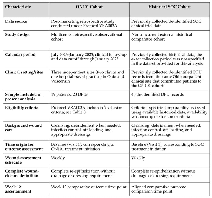
This study was a multicenter retrospective observational analysis conducted in the US to evaluate the clinical effectiveness of ON101 in the management of chronic DFUs. The analysis included patients treated with ON101 in conjunction with standard wound care and an external historical SOC comparator cohort comprising 40 de-identified DFU records derived from previously collected SOC clinical trial data. The historical SOC cohort was nonconcurrent with the ON101 cohort, and treatment allocation between the two cohorts was not randomized within the present study. Cohort flow diagram is shown in Figure 1. The 40 historical SOC records were selected retrospectively by the participating physician after follow-up of the ON101 cohort had occurred. The total number of potentially eligible records in the historical source population was not available for the present analysis, and no contemporaneous screening log or prespecified record-selection algorithm was available. The comparator records therefore cannot be considered a documented consecutive series. Considering that record selection occurred retrospectively, clinical outcomes may have been known at the time of selection; accordingly, potential outcome-informed selection cannot be excluded.
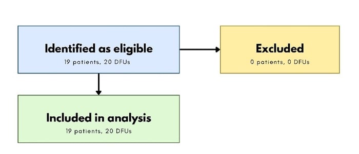
Figure 1. Flow of ON101-treated patients and diabetic foot ulcers into the analytic cohort. No prospective screening log was maintained.
The ON101 cohort was evaluated under a post-marketing retrospective research protocol (VRA003A, Version A, dated January 25, 2024) using clinical data collected during routine wound care. Protocol VRA003A was finalized before retrospective record identification, selection, and data extraction. The cohort included patients treated with ON101 at participating U.S. sites between July 2023 and January 2025, with clinical follow-up available through January 2025. The ON101 cohort included three participating centers, consisting of two clinics and one hospital-based practice, located in Ohio and Wisconsin, USA. The three sites contributed 8 DFUs from 8 patients, 6 DFUs from 6 patients, and 6 DFUs from 5 patients, respectively. Sites were selected as a convenience sample of collaborating clinicians experienced in chronic wound management who used ON101 in routine post-marketing practice and agreed to provide the protocol-specified clinical information. At two sites, clinicians identified potentially eligible records using the protocol-defined criteria; at the third site, records were identified after Oneness provided training on the same eligibility criteria and data-collection requirements. No formal protocol-specific clinical training was provided for ON101 application, wound assessment, or routine patient care; the product training at the third site addressed retrospective case identification and the protocol-defined data requirements. The cases were not identified through a formally documented consecutive screening process; inclusion was based on protocol eligibility and availability of the required clinical documentation. No additional centralized standardization of treatment or wound-assessment procedures was implemented beyond the protocol-defined retrospective data requirements. Photography and wound-area measurement were performed using each site's routine equipment and documentation methods.
Considering that this was a retrospective post-marketing study, potentially eligible cases were identified from existing ON101-treated clinical records rather than through prospective screening, and a separate prospective screening log was not maintained. A total of 19 patients with 20 DFUs were identified as meeting the protocol-defined eligibility criteria and having the clinical documentation required for analysis. No identified eligible ON101-treated cases were subsequently excluded; all 19 patients with 20 DFUs comprised the analytic cohort. Outcome-dependent case selection cannot be completely excluded because wound outcomes may have been known for some retrospectively identified records. The data cutoff for the present analysis was January 2025; no clinical outcomes occurring after this date were included. The historical SOC dataset was not assembled prospectively under Protocol VRA003A, and equivalence of all protocol eligibility criteria could not be established retrospectively. Instead, comparability was evaluated criterion by criterion using variables available in the historical dataset. Criteria for which corresponding historical information was unavailable or incomplete were classified as not verifiable rather than assumed to have been met.
For the ON101 cohort, patient demographics, ulcer characteristics, treatment exposure, wound-bed characteristics, wound-area progression, and wound closure outcomes were assessed where available.
Comparative analyses between the ON101 and historical SOC cohorts were conducted for predefined wound outcomes using variables available in both data sources. Baseline characteristics of the historical SOC cohort were summarized using aggregate-level descriptive statistics in accordance with data-use restrictions, whereas comparative outcome analyses were based on the available de-identified data. Given the nonconcurrent historical comparator design, between-cohort findings were interpreted as exploratory associations rather than causal estimates of treatment effects.
Table 1. Comparison of Key Design and Outcome-Ascertainment Characteristics of the ON101 and Historical SOC Cohorts.
ETHICAL APPROVAL AND PATIENT CONSENT
The study was conducted in accordance with the ethical principles outlined in the Declaration of Helsinki and applicable regulatory requirements for retrospective research involving human subjects. The study protocol (Version A, dated 25 Jan 2024) received approval from the WCG Institutional Review Board (IRB) under Study Number 1372521. The IRB granted a waiver of informed consent under 21 CFR 50.22, as the research involved minimal risk and relied exclusively on previously collected clinical data. All patient data were fully de-identified before analysis to ensure confidentiality and compliance with HIPAA privacy requirements.
STUDY POPULATION
The comparative study population comprised 60 DFUs, including 20 DFUs from 19 patients treated with ON101 and 40 DFU records from the external historical SOC comparator cohort.
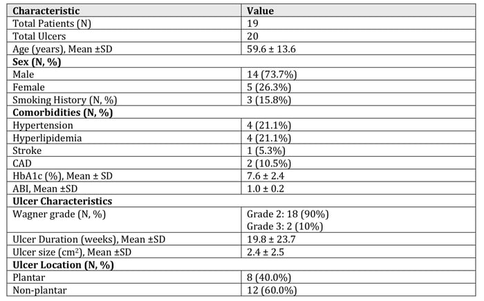
The ON101 cohort included 20 DFUs from 19 patients treated during routine wound care in the US. All patients were adults with diabetes and had ulcers below the malleolus present for at least four weeks before the treatment initiation. The mean (SD) age was 59.6 ± 13.6 years; 14 patients (73.7%) were male and 5 (26.3%) were female. Target ulcers were characterized according to the Wagner grade system, plantar or non- plantar location, and the presence of infection. Detailed patient demographics and clinical characteristics of the ON101 cohort are summarized in Table 2.
Table 2. Baseline Patient and Ulcer Characteristics of the ON101 Cohort.
The external historical SOC comparator cohort comprised 40 de-identified DFU records from a single outpatient clinical site in Ohio. The comparator data were provided for the present analysis in de-identified form and were summarized using aggregate-level descriptive statistics in accordance with applicable data-use restrictions. Mean (±SD) age was 60.6 ± 13.9 years among 36 patients with available age data, and sex was available for all 40 records, of which 28 (70.0%) were male and 12 (30.0%) were female. Baseline clinical characteristics included a mean HbA1c of 8.1 ± 1.8%, a mean baseline wound area of 5.1 ± 4.0 cm², and a mean ulcer duration of 22.2 ± 14.5 weeks among the 23 records with available duration data. These aggregate summaries are provided to characterize the historical comparator cohort without disclosure of individual-level baseline data. No formal between-cohort baseline comparison table was generated.
ELIGIBILITY CRITERIA
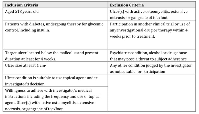
Eligibility criteria for the ON101 cohort are presented in Table 3. For the external historical SOC cohort, comparability with the ON101 eligibility criteria was assessed using the variables available in the historical dataset. Age was available for 36 of 40 historical records, and ulcer duration was available for 23 of 40 records.
Availability of other eligibility-related variables is reported where applicable. Eligibility criteria that could not be verified because the corresponding historical variable was unavailable or incomplete were not assumed to have been met. For efficacy analyses, the protocol-defined evaluable population included patients who received ON101 for at least four weeks, including those who achieved complete ulcer healing at any visit during the treatment period, and who had at least one post-treatment target-ulcer assessment.
Table 3. Inclusion and Exclusion Criteria for the ON101 Cohort.
TREATMENT PROCEDURE
ON101 was applied to the ulcer twice daily to cover the wound surface for up to 16 weeks or until complete wound closure. All patients received SOC for the management of the DFU, including cleansing, debridement when needed, infection control, off-loading, and appropriate dressings. No other topical agents or advanced wound products were used during treatment. Systemic antibiotics were used when clinically indicated. Other topical antibiotics, topical steroids, antiseptic soaps, growth factors, grafts, or investigational treatment were not applied to the target ulcer during the study period.
For the external historical SOC cohort, treatment consisted of standardized wound care treatment, including sharp debridement, offloading, dressings, infection management, and systemic risk factors management, such as glycemic control, comparable to the standard wound care in ON101 cohort.
DATA COLLECTION AND WOUND ASSESSMENT
Clinical data were retrospectively extracted from the medical records of patients and recorded in standardized case report forms (CRFs). The following categories of data were collected:
-
Demographics and medical history
-
Sex, height, body weight
-
Smoking habits
-
Relevant medical history within three months before treatment initiation
-
Clinical assessments
-
Area of the serial ulcer
-
Wound bed characteristics
-
Ulcer duration before treatment
-
Off-loading device usage
-
Photographic documentation of the target ulcer
-
Laboratory and physiological parameters
-
White blood cell count (WBC) and C-reactive protein (CRP) within one week before treatment
-
HbA1c, albumin, creatinine, and hemoglobin within three months before treatment
-
Vascular perfusion assessments using ankle-brachial index (ABI), transcutaneous oxygen measurement (TcPO₂), or Doppler ultrasound
-
Treatment exposure
-
Documentation of concomitant medications
-
Record of prior therapies within four weeks before treatment
-
Outcome Measures
The primary objective of this study was to compare complete wound closure by Week 12 between the ON101-treated cohort and the external historical SOC comparator cohort. For the ON101 cohort, the time origin for outcome assessment was from the first/baseline visit, with wound assessments performed weekly.
Complete wound closure was defined as complete re-epithelization without drainage or dressing requirement assessed by the investigators. For the historical SOC cohort, the corresponding time origin was from the first/baseline visit, with wound assessments performed weekly. For both cohorts, complete wound closure was defined as complete re-epithelialization without drainage or dressing requirement and was assessed by the treating physician/investigator based on routine clinical documentation. No protocol-required confirmatory assessment at a subsequent visit or blinded central adjudication of wound closure was performed. Week 12
was used as the aligned comparative time point for both cohorts.
Week 4 PAR was evaluated as an exploratory endpoint and was calculated from the change in wound area from baseline to the Week 4 assessment. This endpoint was used to quantify early wound-area response before the primary Week 12 closure assessment. An exploratory analysis examined the association between achieving
≥50% PAR at Week 4 and subsequent complete wound closure by Week 12; this analysis was descriptive and was not intended to validate ≥50% PAR as a prognostic threshold in this dataset. Supportive and exploratory endpoints included the proportion of DFUs achieving ≥50% PAR at Week 4, cumulative complete wound closure at Weeks 4, 6, 8, and 12, and time to complete wound closure. Wound-bed characteristics were evaluated as supportive descriptive outcomes and included granulation tissue coverage, slough, eschar, and re-epithelialization where available. Considering that these characteristics were not consistently captured using harmonized definitions and measurement scales across the ON101 and historical SOC datasets, wound-bed findings were not interpreted as independent comparative efficacy outcomes.
Exploratory subgroup analyses examined Week 12 complete wound closure according to ulcer location (plantar versus non-plantar). These analyses were considered hypothesis-generating, and potential effect modification by ulcer location was further assessed using formal interaction tests.
STATISTICAL ANALYSIS
Categorical variables were summarized as counts and percentages, and continuous variables as means with standard deviations (SDs) and medians with interquartile ranges (IQRs). Between-cohort comparisons were performed using Fisher’s exact test for categorical variables and the Mann–Whitney U test for continuous
variables. The DFU was the primary unit of analysis. A total of 20 ON101-treated DFUs were contributed by 19 patients, one patient contributed two DFUs; therefore, complete independence of all ulcer-level observations could not be assumed. Baseline characteristics were summarized descriptively without formal hypothesis
testing because individual-level baseline data were not uniformly available for the external historical SOC cohort.
The primary endpoint was complete wound closure by Week 12. Complete-closure proportions at Weeks 4, 6, 8, and 12 were presented as counts and percentages with Wilson score 95% CIs calculated without continuity correction. Between-cohort differences in closure proportions were assessed using Fisher’s exact
test.
Week 4 PAR was analyzed as an exploratory early-response outcome and was summarized among evaluable wounds using the mean and standard deviation, and the median and interquartile range. Between-cohort distributions were compared using the Mann–Whitney U test. Wounds were considered evaluable if they had either documented complete closure on or before Week 4, or observed baseline and Week 4 wound-area measurements. Documented complete closure on or before Week 4 was assigned a PAR of 100% as an endpoint-derivation rule. Accordingly, Week 4 PAR values did not uniformly represent paired baseline and Week 4 wound-area measurements, because wounds that had already achieved complete closure were assigned a PAR of 100%. The proportion of evaluable DFUs achieving Week 4 PAR ≥50% was compared between cohorts using Fisher’s exact test, with the odds ratio and corresponding 95% confidence interval reported.
Due to the absence of Week 4 PAR data for 10 of the 20 ON101-treated DFUs, a sensitivity analysis was performed in which wounds without evaluable Week 4 PAR were classified as nonresponders for the binary PAR ≥50% endpoint. This rule was applied only to the binary responder analysis; no continuous PAR values
were imputed.
Complete-closure proportions at Weeks 4, 6, and 8 were evaluated as supportive longitudinal outcomes. Time to complete wound closure through Week 12 was analyzed using the Kaplan–Meier method, and the ON101 and historical SOC cohorts were compared using the log-rank test. DFUs without documented closure
by Week 12 were censored at their last available wound assessment or at Week 12, whichever occurred first. Closure documented after Week 12 was administratively censored at Week 12. The cumulative probability of complete closure was presented as 1 − S(t). Kaplan–Meier median time to closure was reported when estimable; otherwise, it was reported as not reached. Observed time to closure among DFUs that achieved closure was summarized separately using the median and IQR.
The prognostic relationship between observed early wound response and subsequent Week 12 closure was explored descriptively within the ON101 cohort. Among DFUs with evaluable Week 4 PAR, the positive predictive value of achieving PAR ≥50% for subsequent complete closure by Week 12 was calculated. A separate sensitivity analysis classified DFUs without evaluable Week 4 PAR as nonresponders and summarized the resulting association using an odds ratio and Fisher’s exact test.
Wound-bed characteristics and ulcer-location subgroup findings were considered supportive or exploratory analyses.
Propensity scores were estimated using baseline wound area and plantar ulcer location. The historical comparator dataset did not contain complete individual-level information for several clinically important prognostic variables, therefore, the propensity-score model was intentionally limited to covariates available with sufficient completeness and overlap across cohorts. Overlap weighting was applied, and covariate balance was assessed using standardized mean differences, with an absolute standardized mean difference of <0.10 indicating acceptable balance. Covariate balance before and after weighting, propensity-score overlap, and effective sample sizes are reported in further datasets that can be provided upon author request.
Effect modification by ulcer location was evaluated using formal interaction tests. For complete wound closure by Week 12, a logistic regression model including treatment group, ulcer location, and a treatment-by-location interaction term was fitted. For time to complete closure, a Cox proportional hazards model containing
the corresponding interaction term was used. Interaction p-values, rather than differences between subgroup-specific p-values, were used to assess heterogeneity of the observed treatment-group association. The Week 12 complete wound closure comparison was the primary analysis. All other analyses were considered
supportive or exploratory. No adjustment for multiplicity was performed; therefore, p-values from secondary, supportive, subgroup, sensitivity, and prognostic analyses were interpreted descriptively and were not considered confirmatory.
No continuous missing values were imputed, and available-case denominators were used for the Week 4 PAR analysis. Documented complete closure on or before Week 4 was assigned a PAR of 100% as an endpoint-derivation rule rather than as imputation. For the binary Week 4 PAR ≥50% sensitivity analysis only, wounds
without evaluable Week 4 PAR were classified as nonresponders. No interpolation or last-observation-carried-forward procedure was applied. All tests were two-sided, with P<0.05 considered statistically significant.
RESULTS
The comparative analysis included 60 DFUs, comprising 20 DFUs treated with ON101 and 40 DFUs from the external historical SOC comparator cohort. Comparative outcomes were assessed through Week 12, with additional descriptive follow-up of the ON101 cohort beyond Week 12 where available.
TREATMENT EXPOSURE AND CONCOMITANT WOUND CARE
Detailed longitudinal information regarding ON101 adherence, cumulative treatment exposure, frequency of debridement, duration of offloading, systemic antibiotic use, temporary treatment interruptions, discontinuations, and other concomitant wound interventions could not be reconstructed reliably and consistently across the retrospective records. These variables were therefore not summarized quantitatively.
PRIMARY ENDPOINT: COMPLETE WOUND CLOSURE AT WEEK 12
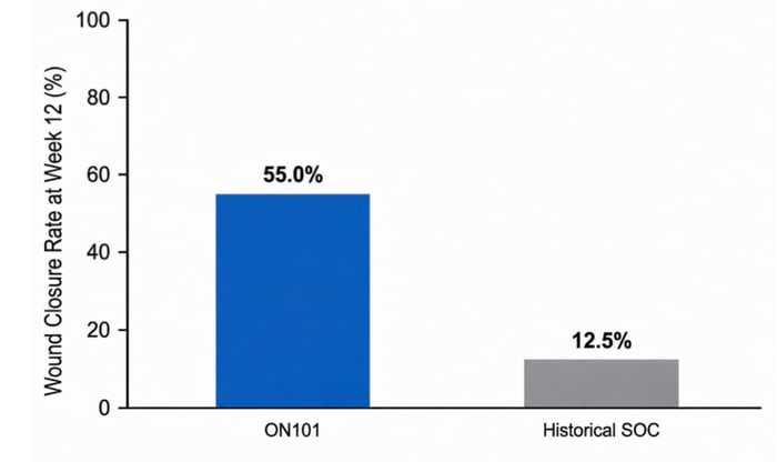
By Week 12, complete wound closure was observed in 11 of 20 DFUs (55.0%) in the ON101 cohort and 5 of 40 DFUs (12.5%) in the historical SOC cohort, corresponding to an absolute risk difference of 42.5 percentage points (95% CI, 17.6–62.9) and a risk ratio of 4.40 (95% CI, 1.77–10.94; Fisher’s exact p=0.0012). The observed Week 12 closure rates are shown in Figure 2.
In an exploratory propensity-score overlap-weighted sensitivity analysis of the full analytic cohort (ON101, n=20; historical SOC, n=40), estimated Week 12 complete closure rates were 55.3% and 12.0%, respectively, corresponding to an adjusted risk difference of 43.2 percentage points (95% bootstrap CI, 10.6– 71.5) and an adjusted risk ratio of 4.59 (95% bootstrap CI, 1.55–26.46). Adequate balance was achieved for both covariates included in the propensity-score model, with post-weighting absolute standardized mean differences <0.10.
Figure 2. Comparison of Complete Wound Closure Rate at Week 12 between the ON101 and Historical
SOC Cohorts.
EXPLORATORY EARLY WOUND-AREA RESPONSE AT WEEK 4
Week 4 PAR was evaluable in 10 of 20 ON101-treated DFUs (50.0%) and all 40 historical SOC DFUs. In the ON101 cohort, three DFUs had documented complete closure before Week 4 and two achieved complete closure at Week 4; these five DFUs were assigned a Week 4 PAR of 100%. The remaining five evaluable ON101- treated DFUs had observed baseline and Week 4 wound-area measurements. Thus, the Week 4 PAR analysis included both PAR values derived from paired wound-area measurements and PAR values assigned as 100% based on documented complete closure by Week 4. Among evaluable DFUs, mean (SD) Week 4 PAR was 86.0%±23.6% in the ON101 cohort and 8.6%±84.0% in the historical SOC cohort. The corresponding medians (IQRs) were 100% (73%–100%) and 27% (−3.75%–55%), respectively (Mann–Whitney U p=0.00006).
Among evaluable DFUs, 9 of 10 ON101-treated DFUs (90.0%) and 13 of 40 historical SOC DFUs (32.5%) achieved Week 4 PAR ≥50% (odds ratio, 18.69; 95% CI, 2.14–163.61; Fisher’s exact p=0.0027). However, Week 4 PAR was evaluable for only 10 of 20 ON101-treated DFUs, and this available-case comparison was therefore considered exploratory and potentially susceptible to selection bias. In a sensitivity analysis classifying the 10 ON101-treated DFUs without evaluable Week 4 PAR as nonresponders, 9 of 20 ON101-treated DFUs (45.0% and 13 of 40 historical SOC DFUs (32.5%) achieved the threshold (odds ratio, 1.70; 95% CI, 0.57–5.11; Fisher’s exact p=0.401). Thus, the apparent between-cohort difference observed in the available-case analysis was attenuated and was not statistically significant under the missing-as-nonresponder assumption, indicating sensitivity to the handling of missing Week 4 assessments.
Among the nine ON101-treated DFUs with an observed Week 4 PAR ≥50%, eight achieved complete closure by Week 12 and one did not, corresponding to a descriptive PPV of 88.9%. However, only one evaluable DFU had an observed Week 4 PAR <50% and this DFUs also achieved complete closure by Week 12.
Accordingly, the limited available-case data did not permit reliable estimation of an association between observed Week 4 responder status and subsequent closure. (Table 4)
Table 4. Association Between Week 4 PAR ≥50% and Complete Wound Closure by Week 12 in the ON101 Cohort.
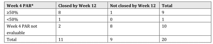
*Week 4 PAR was evaluable in 10 of 20 ON101-treated DFUs. Among the nine DFUs with observed Week 4 PAR ≥50%, eight achieved complete closure by Week 12. Only one evaluable DFU had observed Week 4 PAR <50%; therefore, these data were considered descriptive and were not used to validate ≥50% PAR as a prognostic threshold
LONGITUDINAL COMPLETE WOUND CLOSURE AND TIME-TO-CLOSURE
Differences in wound closure were evident at earlier assessment time points and persisted through Week 12. By Week 4, complete closure had been achieved in 5 of 20 ON101-treated DFUs (25.0%; 95% CI, 11.2%– 46.9%) compared with 1 of 40 historical SOC DFUs (2.5%; 95% CI, 0.4%–12.9%; p=0.0132). By Week 6, the corresponding closure proportions were 40.0% (8/20; 95% CI, 21.9%–61.3%) and 2.5% (1/40; 95% CI, 0.4%– 12.9%), respectively (p=0.0004). At Week 8, 9 of 20 ON101-treated DFUs (45.0%; 95% CI, 25.8%–65.8%) had achieved complete closure compared with 4 of 40 historical SOC DFUs (10.0%; 95% CI, 4.0%–23.1%; p=0.0057). By Week 12, the closure proportions had increased to 55.0% and 12.5%, respectively (p=0.0012).
Among wounds that achieved complete closure during available follow-up, the median observed time from treatment initiation to documented closure was 5.0 weeks (IQR, 3.5–6.0 weeks) in the ON101 cohort and 8.0 weeks (IQR, 8.0–8.0 weeks) in the historical SOC cohort. This analysis was restricted to DFUs with documented closure and was therefore interpreted descriptively. During the available ON101 follow-up through Week 16, complete closure was documented in 15 of 20 DFUs (75.0%; 95% CI, 53.1%–88.8%). This post–Week 12 observation was descriptive and was not compared with the historical SOC cohort (Table 5).
Table 5. Complete Wound Closure and Time-to-Closure Outcomes.
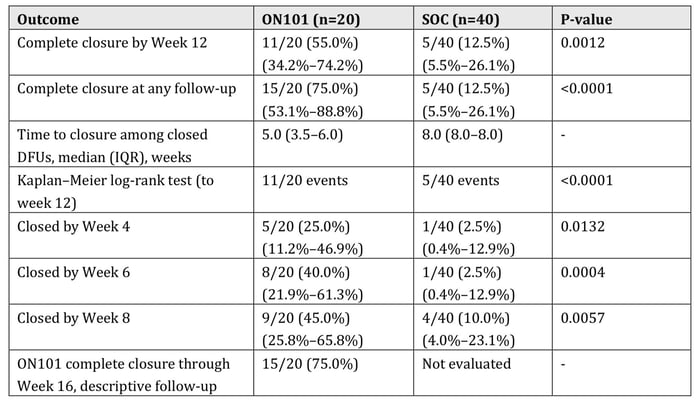
*Post–Week 12 follow-up was descriptive for the ON101 cohort and was not compared with the historical SOC cohort.
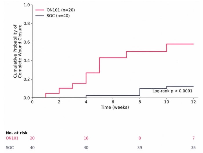
Time-to-event analysis demonstrated separation in the cumulative probability of complete wound closure between the two cohorts over the 12-week observation period (Figure 3). Eleven closure events occurred among 20 ON101-treated DFUs and 5 closure events among 40 historical SOC DFUs by Week 12. Kaplan–Meier curves showed separation between cohorts through Week 12 (log-rank p<0.0001), with earlier accumulation of documented closure events in the ON101 cohort.
Figure 3. Kaplan–Meier estimated cumulative probability of closure through week 12 for the ON101 cohort (n=20) and SOC comparator cohort (n=40). DFUs without documented complete closure were censored at the last available wound assessment or Week 12, whichever occurred first.
Exploratory analyses by ulcer location showed no evidence of effect modification for complete wound closure by Week 12 (treatment-by-location interaction p=0.912) or for time to complete closure (interaction p=0.950). However, these interaction analyses were imprecise because of the small subgroup sizes and low number of closure events, particularly in the plantar historical SOC subgroup. Detailed subgroup results are provided in supplementary datasets which can be provided upon author request.
SUPPORTIVE WOUND-BED PROGRESSION AND CHARACTERISTICS
Wound-bed characteristics were evaluated as supportive descriptive outcomes. At baseline, granulation tissue coverage was available for 10/20 ON101 DFUs and 40/40 historical SOC DFUs. Mean (SD) granulation tissue coverage was 74.5% (39.8%) and 73.8% (9.3%), respectively. Median granulation coverage was 97.5% (IQR, 60%–100%) in the ON101 cohort and 80% (IQR, 70%–80%) in the historical SOC cohort. Granulation coverage below 50% was observed in 3/10 (30.0%) ON101 DFUs and 0/40 (0%) historical SOC DFUs, respectively. During the available ON101 follow-up through Week 16, 14 of 20 DFUs (70.0%) had achieved complete re-epithelialization, while the remaining open wounds showed a descriptive shift toward predominantly granulating tissue with limited residual slough or eschar.
Granulation tissue coverage was available using a comparable quantitative measure in both cohorts and was therefore evaluated between groups. Other wound-bed characteristics, including slough, eschar, and re- epithelialization, were not consistently captured using harmonized definitions or measurement scales across both datasets and were therefore interpreted descriptively without formal between-cohort statistical comparison.
SAFETY AND TOLERABILITY
Adverse events were not systematically ascertained under a prospective safety-surveillance framework in this retrospective study. Available clinical records were reviewed for documented safety information; however, absence of documentation could not be interpreted as absence of an event. Accordingly, no formal conclusions regarding the safety or tolerability of ON101 were drawn from this dataset.
DISCUSSION
The principal finding of this retrospective comparative study was the higher proportion of complete wound closure by Week 12 observed in the ON101 cohort compared with the external historical SOC cohort (55.0% versus 12.5%; p=0.0012). This difference in the definitive clinical outcome was accompanied by progressively higher closure proportions during follow-up and earlier accumulation of complete closure events. Given the nonrandomized and nonconcurrent nature of the historical comparison, however, these findings should be interpreted as comparative associations rather than causal estimates of treatment effect.
Baseline comparability remains an important consideration when interpreting the magnitude of the observed between-cohort differences. ON101-treated DFUs had a smaller mean baseline wound area than DFUs in the historical SOC cohort (2.4 ± 2.5 cm2 vs 5.1 ± 4.0 cm2) and similar baseline granulation tissue coverage (74.5% versus 73.8%), both of which may influence subsequent wound closure. To evaluate the robustness of the primary finding, an exploratory propensity score overlap-weighted sensitivity analysis was conducted in the full analytic cohort using baseline wound area and plantar ulcer location. The adjusted estimates remained directionally consistent with the unadjusted findings, with estimated Week 12 closure rates of 55.3% and 12.0% and an adjusted risk difference of 43.2 percentage points. Considering that the historical comparator dataset did not contain complete individual-level information for several clinically important prognostic variables, the propensity-score model was intentionally limited to covariates available with sufficient completeness and overlap across cohorts. Residual imbalance and unmeasured confounding therefore cannot be excluded, and the propensity-score analysis should be regarded as supportive sensitivity evidence rather than a fully adjusted causal estimate of treatment effect.
Complete wound closure was selected as the primary clinical outcome because it represents definitive wound resolution and provides an intuitive measure of treatment success in DFU care. Importantly, the observed between-cohort difference was not confined to the Week 12 assessment. Complete closure was documented in 25.0% versus 2.5% of DFUs by Week 4, 40.0% versus 2.5% by Week 6, 45.0% versus 10.0% by Week 8, and 55.0% versus 12.5% by Week 12 in the ON101 and historical SOC cohorts, respectively. Kaplan–Meier analysis further demonstrated significant separation in the cumulative probability of complete closure through Week 12 (log-rank p<0.0001). Among DFUs that achieved complete closure during available follow- up, the median observed time to closure was 5.0 weeks in the ON101 cohort and 8.0 weeks in the historical SOC cohort. This latter comparison was restricted to wounds that achieved closure, therefore, it is descriptive rather than a Kaplan–Meier median or a causal estimate of reduction in healing time. Taken together, the serial closure and time-to-event findings indicate that the primary Week 12 result was part of a broader observed pattern of earlier wound resolution rather than an isolated difference at a single assessment.
Week 4 PAR provided exploratory information regarding early wound response before the primary Week 12 closure assessment. Among evaluable wounds, mean Week 4 PAR and the proportion achieving PAR ≥50% were greater in the ON101 cohort than in historical SOC. Interpretation of these findings should also account for the endpoint-derivation rule whereby DFUs documented as completely closed on or before Week 4 were assigned a PAR of 100%; therefore, not all Week 4 PAR values were derived from paired wound-area measurements. However, Week 4 PAR was evaluable in only 10 of the 20 ON101-treated DFUs. The available- case comparison was therefore susceptible to selection bias, and the between-cohort association was substantially attenuated when wounds without evaluable Week 4 PAR were classified as nonresponders. Among the 9 observed ON101 responders, eight subsequently achieved complete closure by Week 12, corresponding to a descriptive PPV of 88.9%; however, only one observed nonresponder was available for comparison. In addition, when ON101-treated DFUs without evaluable Week 4 PAR were classified as nonresponders in a sensitivity analysis, the apparent between-cohort difference in ≥50% PAR was attenuated and was not statistically significant (45.0% vs 32.5%; Fisher’s exact p=0.401). These findings should therefore be regarded as hypothesis-generating and do not validate Week 4 PAR ≥50% as a prognostic or surrogate threshold in this dataset. Accordingly, Week 4 PAR provides complementary evidence of early wound progression rather than as an independent demonstration of comparative efficacy.
Wound-bed observations provided additional supportive clinical context for the wound-area and closure findings. During ON101 treatment, the recorded wound-bed profile showed a descriptive progression toward granulation and re-epithelialization, with 70.0% of DFUs achieving complete epithelialization by the end of 16- week. However, wound-bed characteristics, including slough, eschar, and re-epithelialization, were not consistently captured using harmonized definitions and measurement scales across the ON101 and historical SOC datasets. Accordingly, wound-bed findings were considered supportive descriptive observations and were not interpreted as independent evidence of comparative efficacy.
Exploratory analyses by ulcer location showed similar directional findings for plantar and non-plantar DFUs. Interaction analyses found no evidence that ulcer location modified the observed association between treatment cohort and Week 12 complete closure or time to closure. Nevertheless, the subgroup sample sizes and number of closure events were small, resulting in limited precision. These findings should therefore be interpreted as an absence of detectable effect modification rather than evidence that outcomes were equivalent across ulcer locations.
From a clinical perspective, the combination of a higher fixed-time complete closure rate, progressively greater closure proportions across serial assessments, and earlier accumulation of closure events provides a more complete characterization of wound resolution than any single measure considered in isolation. The exploratory Week 4 PAR findings provide additional information regarding early wound progression but should remain secondary to the more definitive closure outcomes because of incomplete Week 4 assessments. Overall, the findings are consistent with an earlier observed progression toward complete wound resolution in the ON101 cohort relative to the historical SOC comparator. However, the present study did not systematically evaluate hospitalization, amputation, physician visits, subsequent procedures, or healthcare costs across both cohorts. Reductions in these downstream outcomes therefore cannot be inferred from the present analysis, which provides comparative clinical evidence focused specifically on wound progression and resolution.
LIMITATIONS
Several limitations should be considered when interpreting these findings. First, the study used a nonconcurrent external historical SOC comparator rather than a concurrently enrolled randomized controlled cohort. Differences in source-study design, calendar period, patient selection, treatment setting, background wound care, assessment schedules, and endpoint ascertainment may therefore have introduced selection bias, information bias, and residual confounding. Complete wound closure was assessed by the treating physician without required confirmation at a subsequent visit or blinded central adjudication, which may have introduced additional outcome-ascertainment variability.
Second, the ON101 and historical SOC cohorts were not fully comparable at baseline. An exploratory propensity score overlap-weighted sensitivity analysis restricted to Wagner Grade 2 DFUs was used to adjust for baseline wound area and plantar ulcer location, and the adjusted estimates remained directionally consistent with the primary unadjusted analysis. However, incomplete availability of other potentially relevant prognostic variables limited the scope of covariate adjustment. Residual imbalance remained in ulcer duration and baseline granulation tissue coverage, and unmeasured differences associated with the use of a nonconcurrent historical comparator cannot be excluded. Accordingly, the propensity score analysis should be considered supportive sensitivity evidence rather than a fully adjusted causal estimate of treatment effect.
Third, Week 4 PAR was evaluable in only 10 of the 20 ON101-treated DFUs. Wounds with evaluable Week 4 PAR had a higher observed Week 12 closure proportion than wounds without evaluable Week 4 PAR, suggesting that outcome availability may have been related to subsequent healing. Consequently, the available- case PAR analysis may be affected by selection bias and may overestimate the early-response difference between cohorts. In addition, the between-cohort association was markedly attenuated in the missing-as- nonresponder sensitivity analysis. The Week 4 PAR results should therefore be interpreted as exploratory.
Fourth, wound-bed characteristics were not uniformly measured using harmonized definitions across cohorts, limiting formal comparative interpretation of these measures. Finally, the modest sample size, particularly within subgroup analyses, produced imprecise effect estimates and limits generalizability.
Considering that the participating sites were a convenience sample and cases were not identified through a documented consecutive screening process, selection bias and outcome-dependent case selection cannot be excluded. In addition, treatment and assessment procedures were not centrally standardized; site-specific equipment and documentation methods for photography and wound-area measurement may have introduced measurement variability. The small number of DFUs within each site limited the feasibility of formal adjustment for site-level clustering, and residual site-level confounding cannot be excluded. In addition, one patient contributed two target DFUs. Analyses were conducted at the ulcer level, therefore, the assumption of independence between observations may not have been fully met, and potential within-patient correlation could have affected the precision of the inferential estimates. Accordingly, these estimates should be interpreted cautiously. These limitations support interpretation of the between-cohort findings as exploratory comparative evidence rather than causal estimates of treatment superiority.
Despite these limitations, the concordance of the primary Week 12 closure outcome with early Week 4 wound-area reduction, serial closure assessments, and time-to-event findings provides a consistent clinical signal across complementary measures of wound progression. The results support further prospective evaluation of ON101 within controlled comparative studies designed to account for baseline wound severity and wound-bed characteristics and to determine whether the observed earlier progression toward complete closure is reproducible in larger and more diverse DFU populations.
CONCLUSION
In this retrospective comparative study, ON101 plus standard wound care was associated with a higher observed Week 12 complete wound closure rate and earlier accumulation of complete closure events than external historical SOC. Exploratory available-case analyses suggested greater early wound-area reduction with ON101, although these findings were sensitive to missing Week 4 assessments. Given the nonconcurrent historical comparator design and clinically relevant baseline differences between cohorts, the findings should be interpreted as exploratory comparative associations and warrant confirmation in prospective controlled studies.
Author Contributions: Conceptualization, B.A.L.; methodology, B.A.L.; writing-original draft preparation, B.N. and K.G.; writing-review and editing, T.S., B.T. and B.N.; supervision, T.S.; project administration, B.T. All authors have read and agreed to the published version of the manuscript.
Received: 20 August 2026 | Accepted: 30 August 2026
Funding: This research was funded by Oneness Biotech Co., Ltd., but they had no other involvement in the study.
Institutional Review Board Statement: The study was approved by the WCG Institutional Review Board (#1372521 on 25 Jan 2024).
Informed Consent Statement: Patient consent was waived due to the research involved minimal risk and relied exclusively on previously collected clinical data.
Data Availability Statement: The de-identified ON101 analytic dataset may be available from the corresponding author upon reasonable request, subject to applicable institutional and data-protection requirements. The historical SOC data were provided subject to data-use restrictions and are not available for redistribution by the authors. Derived aggregate data supporting the analyses and the statistical analysis code may be shared upon reasonable request to the extent permitted by the applicable data-use arrangements.
Declaration of Generative AI: During the preparation of this work the authors used OpenAI to support drafting and language refinement of portions of this manuscript, including improving clarity, grammar, and flow. The AI tool was not used for data generation, data analysis, interpretation of results, or drawing scientific conclusions. After using this tool/service, the authors reviewed and edited the content as needed and take full responsibility for the content of the publication.
Conflicts of Interest: Thomas Serena, MD serves on the Board of Directors of the Journal of Chronic Wound Care Management & Research. SerenaGroup, Inc. received compensation from Oneness Biotech Co., Ltd. for medical writing and editorial support related to preparation of this manuscript.
Disclaimer/Publisher’s Note: The statements, opinions and data contained in all publications are solely those of the individual author(s) and contributor(s) and not of CWRM and/or the editor(s). CWRM and/or the editor(s) disclaim responsibility for any injury to people or property resulting from any ideas, methods, instructions or products referred to in the content.
REFERENCES
1. Hossain MJ, Al-Mamun M, Islam MR. Diabetes mellitus, the fastest growing global public health concern: Early detection should be focused. Health Sci Rep. 2024; 7(3):e2004. https://doi.org/10.1002/hsr2.2004
2. Pan C, Cao B, Fang H et al. Global burden of diabetes mellitus 1990–2021: epidemiological trends, geospatial disparities, and risk factor dynamics. Front Endocrinol. 2025; 16: 1596127 https://doi.org/10.3389/fendo.2025.1596127
3. Parker ED, Lin J, Mahoney T et al. Economic Costs of Diabetes in the U.S. in 2022. Diabetes Care. 2024; 47(1):26–43. https://doi.org/10.2337/dci23-0085
4. Rowley WR, Bezold C, Arikan Y et al. Diabetes 2030: insights from yesterday, today, and future trends. Popul Health Manag. 2017; 20(1):6–12. https://doi.org/10.1089/pop.2015.0181
5. Waibel FWA, Uçkay I, Soldevila-Boixader L et al. Current knowledge of morbidities and direct costs related to diabetic foot disorders: a literature review. Front Endocrinol. 2024; 14:1323315 https://doi.org/10.3389/fendo.2023.1323315=§1
6. Wang R, Gu S, Kim YH et al. Diabetic Wound Repair: From Mechanism to Therapeutic Opportunities. MedComm (2020). 2025; 6(10):e70406. https://doi.org/10.1002/mco2.70406
7. Dawi J, Tumanyan K, Tomas K, Misakyan Y, Gargaloyan A, Gonzalez E, et al. Diabetic Foot Ulcers: Pathophysiology, Immune Dysregulation, and Emerging Therapeutic Strategies. Biomedicines. 2025; 13(5):1076. https://doi.org/10.3390/biomedicines13051076
8. Akkus G, Sert M. Diabetic foot ulcers: A devastating complication of diabetes mellitus continues non-stop in spite of new medical treatment modalities. World J Diabetes. 2022; 13(12):1106–21. https://doi.org/10.4239/wjd.v13.i12.1106
9. Srinivas-Shankar U, Kimyaghalam A, Bergman R. Diabetic Foot Ulceration and Complications. In: StatPearls [Internet]. Treasure Island (FL): StatPearls Publishing; 2026. Available from: http://www.ncbi.nlm.nih.gov/books/NBK499887/
10. Parveen K, Hussain MA, Anwar S et al. Comprehensive review on diabetic foot ulcers and neuropathy: Treatment, prevention and management. World J Diabetes. 2025; 16(3):100329. https://doi.org/10.4239/wjd.v16.i3.100329
11. Ozgok Kangal MK, Kopitnik NL. Physiology, Wound Healing. In: StatPearls [Internet]. Treasure Island (FL): StatPearls Publishing; 2026. http://www.ncbi.nlm.nih.gov/books/NBK535406/
12. Landén NX, Li D, Ståhle M. Transition from inflammation to proliferation: a critical step during wound healing. Cell Mol Life Sci. 2016; 73(20):3861–85. https://doi.org/10.1007/s00018-016-2268-0
13. Xue M, Jackson CJ. Extracellular Matrix Reorganization During Wound Healing and Its Impact on Abnormal Scarring. Adv Wound Care (New Rochelle). 2015; 4(3):119–36. https://doi.org/10.1089/wound.2013.0485
14. Everett E, Mathioudakis N. Update on management of diabetic foot ulcers. Ann N Y Acad Sci. 2018; 1411(1):153–65. https://doi.org/10.1111/nyas.13569
15. Kavitha KV, Tiwari S, Purandare VB et al. Choice of wound care in diabetic foot ulcer: A practical approach. World J Diabetes. 2014; 5(4):546–56. https://doi.org/10.4239/wjd.v5.i4.546
16. Schultz GS, Barillo DJ, Mozingo DW, Chin GA. Wound bed preparation and a brief history of TIME. Int Wound J. 2004; 1(1):19–32. https://doi.org/10.1111/j.1742-481x.2004.00008.x
17. Raja JM, Maturana MA, Kayali S et al. Diabetic foot ulcer: A comprehensive review of pathophysiology and management modalities. World J Clin Cases. 2023; 11(8):1684–93. https://doi.org/10.12998/wjcc.v11.i8.1684
18. Caporusso J, Motley T, Lantis JC, Heisler S, Hicks A, Wu SC, et al. A Multi-Centre, Randomised, Controlled Clinical Trial Assessing Cryopreserved Ultra-Thick Human Amniotic Membrane in the Treatment of Complex Diabetic Foot Ulcers.
Wound Repair Regen. 2025; 33(6):e70110. https://doi.org/10.1111/wrr.70110
19. Boulton AJM, Armstrong DG, Löndahl M et al. New Evidence-Based Therapies for Complex Diabetic Foot Wounds [Internet]. Arlington (VA): American Diabetes Association; 2022 (ADA Clinical Compendia Series). Available from: http://www.ncbi.nlm.nih.gov/books/NBK581559/
20. Fife CE, Eckert KA, Carter MJ. Publicly Reported Wound Healing Rates: The Fantasy and the Reality. Adv Wound Care. 2018; 7(3):77–94. https://doi.org/10.1089/wound.2017.0743
21. Armstrong DG, Swerdlow MA, Armstrong AA et al. Five-year mortality and direct costs of care for people with diabetic foot complications are comparable to cancer. J Foot Ankle Res. 2020; 13(1):16. https://doi.org/10.1186/s13047-020-00383-2
22. Monami M, Ragghianti B, Nreu B et al. Major amputation in non-healing ulcers: outcomes and economic issues. Data from a cohort of patients with diabetic foot ulcers. Int J Low Extrem Wounds. 2025; 24(1):235–244. https://doi.org/10.1177/15347346221097283
23. Mat Saad AZ, Khoo TL, Halim AS. Wound Bed Preparation for Chronic Diabetic Foot Ulcers. ISRN Endocrinol. 2013; 13;2013:608313. https://doi.org/10.1155/2013/608313
24. Zhang Y, Cramb SM, McPhail SM et al. The incidence of and risk factors for hospitalisations and amputations for people with diabetes-related foot ulcers in Queensland, 2011–19: an observational cohort study. Med J Aust. 2025; 223(3):149–58. https://doi.org/10.5694/mja2.52703
25. Nuutila K, Eriksson E. Moist Wound Healing with Commonly Available Dressings. Adv Wound Care. 2021; 10(12):685–98. https://doi.org/10.1089/wound.2020.1232
26. Jin SG. Recent Applications of Chitosan-Based Biomaterials as Wound Dressings. Int J Mol Sci. 2026; 27(4):1637. https://doi.org/10.3390/ijms27041637
27. 28. Product - Bonvadis® [Internet] https://bonvadis.com/us-en/product-us-en/. 2025 May 16 Eriksson E, Liu PY, Schultz GS et al. Chronic wounds: Treatment consensus. Wound Repair Regen. 2022; 30(2):156–71. https://doi.org/10.1111/wrr.12994
29. Halim AS, Khoo TL, Saad AZM. Wound bed preparation from a clinical perspective. Indian J Plast Surg. 2012; 45(2):193– 202. https://doi.org/10.4103/0970-0358.101277
30. McCallon SK, Weir D, Lantis JC. Optimizing Wound Bed Preparation With Collagenase Enzymatic Debridement. J Am Coll Clin Wound Spec. 2015; 6(1–2):14–23. https://doi.org/10.1016/j.jccw.2015.08.003
31. Liu YF, Ni PW, Huang Y, Xie T. Therapeutic strategies for chronic wound infection. Chin J Traumatol. 2022; 25(1):11–6. https://doi.org/10.1016/j.cjtee.2021.07.004
32. Harries RL, Bosanquet DC, Harding KG. Wound bed preparation: TIME for an update. Int Wound J. 2016 22;13(Suppl 3):8–14. https://doi.org/10.1111/iwj.12662
33. Gao J, Moran E, Grimm R et al. The Effect of Primary Care Visits on Total Patient Care Cost: Evidence From the Veterans Health Administration. J Prim Care Community Health. 2022; 13. https://doi.org/10.1177/21501319221141792
34. Riaz M, Iqbal MZ, Klar AS, Biedermann T. Immunomodulatory Mechanisms of Chronic Wound Healing: Translational and Clinical Relevance. MedComm. 2025; 6(11):e70378. https://doi.org/10.1002/mco2.70378
35. Fakher S, Westenberg D. A Comprehensive Overview of Chronic Wound Infections and Current Treatment Methods. Wound Repair Regen. 2025; 33(6):e70115. https://doi.org/10.1111/wrr.70115