
Abstract
Background: Diabetic foot ulcers (DFUs) remain one of the most persistent and debilitating complications associated with diabetes, resulting in significant adverse health outcomes, reduced quality of life, and substantial healthcare costs. Although treatment options have improved, many ulcers still do not fully close under the current standard of care (SOC). These challenges highlight the ongoing need for therapies that are both effective and economically sustainable.
Methods: This randomized controlled multicenter clinical trial assessed whether supplementing SOC with single-layer amniotic membrane (SLAM) improved healing outcomes compared with SOC alone in patients with chronic, non-healing DFUs. The primary outcome was complete ulcer closure over a 12-week treatment period. Wounds were considered closed when full re-epithelialization without drainage was observed at two consecutive weekly assessments (with a ±3-day window), verified by an independent, blinded review panel. Additional analyses included weekly evaluations of percent area reduction (PAR) from TV-1 to TV-13 using both digital planimetry and clinical examination.
Results: In the intention-to-treat (ITT) cohort, 17.9% (n=12/67) of participants receiving SLAM plus SOC achieved full closure, compared with 11.3% (n=7/62) in the SOC group; a higher difference favoring SLAM plus SOC was observed in the per-protocol (PP) population (31.2% SLAM plus SOC [n=10/32], 21.4% SOC [n=6/28]). PAR analyses further indicated that the SLAM-treated group experienced a greater median reduction in wound size over the study period in both ITT and PP populations.
Conclusion: Findings from this interim analysis indicate that adjunctive treatment with SLAM showed a numerical trend toward clinical benefit for patients with chronic DFUs. The consistent trend toward improved closure and enhanced wound area reduction suggests that SLAM could represent a valuable addition to SOC, warranting continued investigation in larger and longer-term studies. The interim findings presented are preliminary, and efficacy requires confirmation in the final analysis.
Keywords: CAMPX; clinical trial design; diabetic foot ulcer; interim analysis; tissue regeneration; xwrap
1 SerenaGroup, Inc., Norwell, MA 02061, USA
* Correspondence: btramelli@serenagroups.com
Diabetic foot ulcers (DFUs) are among the most severe and debilitating complications of diabetes. Their development is multifactorial, resulting from sustained plantar pressure, inappropriate footwear, altered gait mechanics, and repeated mechanical stress that together compromise tissue integrity.1 Underlying pathophysiologic contributors, including peripheral neuropathy, peripheral arterial disease, and metabolic dysregulation, further impair protective sensation, tissue perfusion, immune response, and wound healing capacity, increasing susceptibility to ulcer formation even after minor trauma.2 Clinical assessment and classification systems for DFUs commonly consider wound size and depth, the presence or absence of infection, vascular perfusion, neuropathy, and the ulcer’s anatomical location.3
On a global scale, DFUs occur in an estimated 19–34% of people living with diabetes and are linked to alarmingly high mortality, with reported five-year death rates ranging from 50% to 70%.1 DFUs are also a leading cause of diabetes-related hospitalization and account for a substantial proportion of non-traumatic lower-extremity amputations worldwide, particularly in patients with advanced infection or ischemia.4 Although evidence-based treatment strategies are well-established, healing outcomes remain inadequate for many patients. The current standard of care (SOC), typically consisting of routine sharp debridement, pressure offloading, control of bioburden, and preservation of a moist wound environment, results in complete wound closure within 12 weeks in fewer than half of treated individuals.5
Efforts to prevent DFUs, including regular foot examinations and coordinated care involving both primary care providers and specialists, are critical to lowering incidence rates but often require substantial resources and financial investment.6 An evaluation of the economic impact of DFUs in the United States from 2007 to 2011 demonstrated that healthcare costs for patients with DFUs were approximately $11,000–$17,000 higher per person than for individuals with diabetes without foot ulcers.7 These excess costs are largely attributable to prolonged treatment courses, advanced wound therapies, frequent clinic visits, hospital admissions, and management of complications such as infection and amputation.7
In response to the limitations of conventional therapy, advanced wound care strategies increasingly incorporate cellular, acellular, and matrix-like products (CAMPs). These products are broadly characterized as biomaterials–synthetic or biosynthetic–that promote tissue repair or regeneration through multiple mechanisms of action.8 The use of CAMPs offers several clinical advantages, including stabilization of the wound microenvironment, coverage of exposed anatomical structures, support for surgical wound closure, and improvements in both functional recovery and aesthetic outcomes.8
Previous clinical studies have reported improved healing outcomes in DFUs treated with amniotic membrane-based products when used in conjunction with standard care, although results have varied across products, processing methods, and study designs.9,10 Consequently, additional randomized controlled trials remain necessary to better define the effectiveness of specific amniotic membrane technologies within modern wound care practices.
MATERIALS AND METHODS
CAMPX is a randomized controlled multicenter clinical trial which is evaluating the efficacy of a single layer amniotic membrane (SLAM; XWRAP, Applied Biologics, LLC, US) used as an adjunct to SOC compared with SOC alone for the management of non-healing DFUs (ClinicalTrials.gov identifier #NCT06564831). The interim dataset included 236 patients with non-healing DFUs who were assessed for eligibility at 19 SerenaGroup®, Inc., or affiliated centers in the US. Enrollment began in April 2024, and interim analysis was performed in December 2025. Eligible participants were recruited from individuals receiving care for chronic wounds at outpatient wound clinics.
OBJECTIVES AND ENDPOINTS
The primary objective of the CAMPX clinical trial was to determine the efficacy of SLAM plus SOC versus SOC alone in achieving complete closure of non-healing DFUs over 12 weeks. The primary endpoint was the percentage of target ulcers achieving complete wound closure in 12 weeks.
An additional endpoint evaluated was percentage wound area reduction from TV-1 to TV-13 measured weekly with digital photographic planimetry and physical examination by clinical research coordinators and site investigators, respectively.
Other secondary and exploratory endpoints are deferred to the final analysis.
DIAGNOSIS
The diagnosis of DFUs is primarily established through clinical evaluation, incorporating a comprehensive patient history, physical examination, and selective diagnostic testing. DFUs most frequently develop in areas of the foot that are subjected to repetitive pressure and are commonly associated with peripheral neuropathy and peripheral arterial disease (PAD).
Neuropathic ulcers frequently present with a punched-out appearance, a callused border, and variable granulation tissue. In contrast, ischemic and neuroischemic ulcers often present with irregular wound margins, pale or necrotic wound beds, and minimal exudate. Sensory loss due to neuropathy often results in minimal or absent pain; however, discomfort may be more apparent in the presence of ischemia or superimposed infection.
A thorough medical history is essential for differentiating DFUs from other chronic lower-extremity wounds. Key elements include diabetes duration and control, history of ulceration or amputation, known vascular disease, symptoms of neuropathy, mechanical or traumatic precipitating factors, prior wound management strategies, and footwear practices. Alternative etiologies requiring consideration include venous stasis ulcers, non-diabetic arterial ulcers, pressure injuries, inflammatory or vasculitic ulcers, and malignant lesions.
Neurologic assessment was conducted to evaluate loss of sensation consistent with peripheral neuropathy. Vascular status was assessed in all potential participants, with the ankle-brachial index (ABI) serving as the primary screening tool. Individuals with ABI values between 0.7 and 1.3 were considered eligible, while values >1.3 prompted additional vascular evaluation. In cases where arterial calcification resulted in non-compressible vessels, alternative perfusion assessments such as the toe-brachial index (TBI) were used, with values of ≥0.6 indicating adequate perfusion. Transcutaneous oxygen measurement (TCOM) was also used, and readings of ≥40 mmHg were interpreted as evidence of sufficient tissue oxygenation for study inclusion.
VULNERABLE POPULATIONS
Although vulnerable populations were not specifically recruited, individuals from vulnerable populations may have been present in the potential participant pool
PRODUCT DESCRIPTION
This study assessed a single-layer amniotic membrane (SLAM; XWRAP®, Applied Biologics, LLC, US), a human-derived amniotic membrane allograft intended to function as a protective wound covering for DFUs, venous leg ulcers (VLUs), and pressure ulcers. The product is processed using a technique that preserves the structural integrity of the epithelial membrane, and all donor material undergoes stringent screening and testing in a Clinical Laboratory Improvement Amendments (CLIA)-certified laboratory meeting or exceeding American Association of Tissue Banks (AATB) standards. SLAM is regulated under FDA 21 CFR 1271 and AATB Standards, with processing performed in an FDA-registered, AATB-accredited facility.
Each graft is provided in sterile packaging for single-patient, single-use application, and includes a Tissue Utilization Record with chart labels.
SUBJECT CHARACTERISTICS
Individuals with chronic DFUs were enrolled from participating wound care clinics. After agreeing to follow the study visit schedule, patients reviewed the Institutional Review Board (IRB)-approved consent form. and provided written informed consent. Subsequently, potential participants underwent a screening evaluation to assess eligibility according to the predefined inclusion and exclusion criteria outlined in Table 1.
RANDOMIZATION AND BLINDING
Participants were randomized in a 1:1 ratio using a centralized allocation system and stratified by baseline wound size, wound duration, and patient age. The allocation sequence was generated and maintained within the centralized allocation system; site investigators enrolled eligible participants and obtained treatment assignments through the system after eligibility confirmation. Due to the ongoing enrollment, this report represents a planned interim analysis, so detailed operational characteristics of the randomization schedule are not reported to preserve study integrity.
Due to the nature of the intervention, neither participants nor treating investigators were blinded to treatment assignment. Outcome assessments, wound imaging procedures, and statistical analyses were conducted according to predefined protocol procedures and a pre-specified statistical analysis plan to minimize potential bias.
STUDY PROCEDURES
Participants followed a schedule of study visits that included screening, treatment, wound closure assessment, and post-closure follow-up. This visit framework was designed to support confirmation of eligibility, standardized delivery of wound care, consistent application of investigational and control interventions, and reliable measurement of study endpoints. Investigators and study personnel were trained on protocol-required study procedures prior to study participation, including subject screening and eligibility assessment, informed consent procedures, wound assessment and measurement, wound imaging and fluorescence imaging, debridement procedures, study product application, SOC management, offloading requirements, adverse event reporting, and study documentation requirements. Source documentation, photographic records, treatment records, and electronic case report form data were reviewed as part of routine study monitoring to promote protocol adherence, and data quality across participating sites.
Table 1. Inclusion and Exclusion Criteria.
| Inclusion Criteria | Exclusion Criteria |
|---|---|
| Must be at least 18 years of age. | Known to have a life expectancy of < 6 months. |
| Must have diagnosis of type 1 or type 2 diabetes mellitus. | Target ulcer is not secondary to diabetes. |
| At enrollment, target ulcer with a minimum surface area of 0.7 cm2 and a maximum surface area of 5.0 cm2 measured post debridement using an imaging device. | Target ulcer is infected, requires systemic antibiotic therapy, or there is cellulitis in the surrounding skin. |
| Must have been present for a minimum of 4 weeks and a maximum of 52 weeks of SOC, before the initial screening visit. | Target ulcer exposes tendon or bone. |
| Must be located on the foot with at least 50% of the ulcer below the malleolus. | Evidence of osteomyelitis complicating the target ulcer. |
| Must be Wagner grade 1 or 2, extending at least through the dermis or subcutaneous tissue and may involve muscle, provided it is below the medial aspect of the malleolus. The ulcer may not include exposed tendon or bone. | Is receiving immunosuppressants, including systemic corticosteroids at doses >10 mg of prednisone per day or equivalent, is receiving cytotoxic chemotherapy; or is taking medications that the principal investigator believes may interfere with wound healing (e.g., biologics). |
|
Affected limb must have adequate perfusion confirmed by vascular assessment. Any of the following methods performed within 3 months of the first screening visit are acceptable:
|
Has applied topical steroids to the ulcer surface within one month of initial screening. |
| If the potential subject has two or more ulcers, they must be separated by ≥2 cm. The largest ulcer satisfying the inclusion and exclusion criteria will be designated as the target ulcer. | Has a previous partial amputation on the affected foot that results in a deformity that impedes proper offloading of the target ulcer. |
| Must consent to using the prescribed offloading method for the duration of the study. | Has glycated hemoglobin (HbA1c) ≥12% within 3 months of the initial screening visit. |
| Must agree to attend the weekly study visits required by the protocol. | Surface area of the target ulcer has reduced in size by more than 20% in the 2 weeks prior to the initial screening visit. |
| Must be willing and able to participate in the informed consent process. | The surface area measurement of the target ulcer decreases by ≥20% during the active 2-week screening phase: historical run-in period is the 2 weeks before the initial screening visit (SV); screening phase is 2 weeks between SV and the TV-1 visit during which time the potential subject received SOC. |
| Has an acute Charcot foot, or an inactive Charcot foot, which impedes proper offloading of the target ulcer. | |
| Pregnant or considering becoming pregnant within the next 6 months. | |
| Has end-stage renal disease requiring dialysis. | |
| Participation in a clinical trial involving treatment with an investigational product within the previous 30 days. | |
| Has a medical or psychological condition that may interfere with study assessments. | |
| Treated with hyperbaric oxygen therapy (HBOT) or a Cellular, Acellular, Matrix-like Product (CAMP) in the 30 days prior to the initial screening visit. | |
| Has a malnutrition indicator score <17 as measured on the Mini Nutritional Assessment. | |
| Has a disorder that would create unacceptable risk of post-operative complications. |
SLAM Application Procedures
Subjects randomized to the SLAM plus SOC arm received weekly applications of SLAM during scheduled treatment visits for up to 12 weeks (TV-1 through TV-12) or until complete wound closure was achieved, for a maximum of 12 applications. Prior to each application, the target ulcer was cleaned with normal sterile saline (NSS) and underwent SOC procedures, including cleansing, debridement, moisture balance, and offloading. SLAM was applied directly to the prepared wound bed after cleansing and debridement according to the manufacturer’s instructions for use and covered with a provided calcium alginate or foam dressing. No alternative primary dressings were permitted during the treatment phase. For highly exudative wounds, additional absorptive dressings could be added with approval from the medical monitor. Subjects were permitted one additional unscheduled visit per week for dressing changes when clinically necessary; however, the study product was not routinely reapplied during these visits.
Standard of Care Procedures
All participants received a standardized SOC regimen throughout the screening and treatment phases. SOC consisted of wound cleansing, sharp debridement, moisture management, and pressure offloading.
Wound cleansing was performed using NSS. During the screening phase, antiseptic agents were permitted; however, during the treatment phase, antiseptic agents were limited to wounds demonstrating positive bacterial fluorescence imaging findings or when clinically indicated. Sharp debridement was the preferred debridement modality and was performed to remove necrotic tissue, slough, biofilm, and nonviable tissue. The target ulcer and surrounding skin were prepared with saline or water, and nonviable tissue was excised using sterile instruments. Alternative debridement methods were not permitted during the treatment phase.
Moisture balance was standardized using study-provided calcium alginate or foam dressings. Other primary dressings were prohibited unless specifically approved by the medical monitor. For wounds with excessive exudate, supplemental absorptive dressings were permitted with medical monitor approval.
Offloading was required for plantar ulcers and ulcers exposed to repetitive pressure. The Foot Defender (Defender Ops, US) removable walker boot was the preferred offloading modality across all sites. Subjects unable to tolerate the Defender boot could be treated with total contact cast (TCC), fixed ankle walker, or similar device with approval from the medical monitor. Shoe inserts, half-shoes, and other non-protocol offloading methods were not permitted. Subjects were required to agree to prescribed offloading throughout study participation.
Infection management was standardized across study sites. Subjects with infected target ulcers, cellulitis, osteomyelitis, or infections requiring systemic antibiotic therapy were excluded from enrollment. During the treatment phase, wounds were evaluated at each visit for evidence of infection, including assessment using fluorescence imaging. When infection developed after randomization, treatment was provided according to the investigator’s clinical judgment using topical antimicrobial therapy and/or systemic antibiotics. During active infection, study product application was withheld, dressings were changed at least every 72 hours, and offloading devices were temporarily discontinued until infection resolution.
Adherence to study procedures was assessed throughout the trial. Offloading adherence was evaluated at each treatment visit for subjects not managed with TCC. Participants were educated regarding proper use of offloading devices and maintenance of dressings. Repeated non-adherence was reviewed by the principal investigator and medical monitor and could result in study withdrawal. Completion of at least eight scheduled visits was considered protocol-adherent.
Prohibited therapies during the trial included hyperbaric oxygen therapy (HBOT), additional cellular and/or tissue products (CTPs/CAMPs), enzymatic debridement during the treatment phase, non-study primary dressings without medical monitor approval, and medications judged by the investigator to interfere with wound healing.
Visit Procedures
At the screening visit, written informed consent was obtained prior to initiation of any study procedures. A comprehensive review of medical history was conducted to confirm eligibility. Demographic information, current and prior medical conditions, and concomitant medications were documented. Vascular status was assessed unless qualifying vascular studies had been completed within the preceding three months. Vital signs and a general physical examination were performed. Additional evaluations included nutritional status using the Mini Nutritional Assessment (MNA), documentation of glycated hemoglobin (HbA1c) values (with results from the previous 3 months accepted when available), Wagner ulcer classification, Fitzpatrick skin type assessment, and baseline pain evaluation using the Pain, Enjoyment of Life, General Activity (PEG) scale.
Baseline wound assessment included characterization of granulation tissue, devitalized tissue, wound depth, exudate level, and periwound skin condition. Wound measurements obtained during the two weeks prior to screening were recorded to establish a historical run-in period. Ulcers demonstrating a >20% reduction in surface area during this period were deemed ineligible. Following debridement, each target ulcer was photographed and measured using a standardized imaging system. Fluorescence imaging was also performed to identify bacterial fluorescence within the wound bed or surrounding tissue. Wound photographs and post-debridement surface-area measurements were obtained using the MolecuLightDX or MolecuLight i:X imaging platform (MolecuLight Corp., Canada). Both models were used across sites according to device availability. Newly acquired devices were calibrated by the manufacturer before initial distribution. Manufacturer guidance for the i:X specifies periodic self-diagnosis and does not require user-performed calibration. Before previously used devices were redistributed by the contract research organization, staff completed a manufacturer-supported reset procedure. Any required software updates were installed, the date and time were confirmed, and prior images and user profiles were removed through the device’s data-management functions before the device was reset. Measurement and fluorescence functions were tested before shipment. Fluorescence images were acquired under adequate darkness and at the required distance. For closure adjudication, de-identified images from the closure and closure-confirmation visits were independently assessed by two wound-care specialists who were unaffiliated with the trial and blinded to treatment allocation; when their assessments differed, the determination concordant with a third independent medical lead’s assessment was used.
Throughout the 14-day screening phase, all participants received standardized SOC as described in the Standard of Care Procedures section.
Approximately 14 days following the screening visit, subjects returned for the enrollment visit, at which eligibility criteria were reconfirmed. Adverse events were reviewed, medication use was updated, and a focused physical examination was conducted. Vital signs were obtained, and wound characteristics were reassessed. The percentage area reduction (PAR) observed during the screening period was reviewed and required to remain <20% for continued participation. After debridement, wounds were again photographed and measured using the imaging system. Pain and quality-of-life assessments were administered using the PEG, Forgotten Wound Score (FWS), and wound-specific Quality of Life (wQOL) questionnaires. Fluorescence imaging was repeated. Participants who met all eligibility criteria were electronically randomized and treated according to their assigned study group. SOC procedures were performed. Adherence to offloading protocols was assessed in participants not managed with TCC.
Participants attended weekly treatment visits through week 12 or until complete wound closure, whichever occurred first. At each visit, adverse events and concomitant medications were reviewed, and vital signs were recorded. Wound assessments included evaluation of tissue composition, depth, exudate, and periwound skin condition. Following debridement, wounds were photographed and measured using the imaging system. Pain and quality of life were reassessed using the PEG, wQOL, and FWS questionnaires. Fluorescence imaging was repeated at each visit, and protease swab samples were collected at visits TV-5 and TV-9. SOC procedures were performed at each visit, followed by post-debridement fluorescence imaging. Ulcers were treated according to randomized assignment (SLAM plus SOC or SOC alone) and calcium alginate or foam dressings were applied.
At the final treatment visit (week 13), or earlier if wound closure occurred prior to week 12, adverse events and medication use were reviewed, and pain and quality-of-life assessments were repeated. For ulcers that remained open, detailed wound evaluations were performed, including fluorescence imaging and surface area measurement. Protease swabs were collected, and participants with non-healed wounds were referred for continued wound management. No study product was applied, regardless of whether the ulcer closed or remained open. End-of-study documentation was completed for all participants.
A Closure Confirmation Visit (CCV) was conducted two weeks (±3 days) following the initial determination of complete re-epithelialization, regardless of whether closure was first observed at the final study visit. At this visit, investigators reviewed adverse events and concomitant medications, reassessed pain status, and confirmed wound closure using the imaging system. Complete closure was verified and documented.
Participants who withdrew or were withdrawn prior to study completion underwent procedures equivalent to those performed at the final treatment visit. Unscheduled visits, including those for additional dressing changes, were recorded in the case report form (CRF) with documentation of visit rationale. During such visits, adverse events and medication use were reviewed, and secondary dressings were replaced as necessary.
Upon study exit, participants with unresolved ulcers returned to their treating clinicians for continued SOC management. No additional protocol-mandated follow-up or therapy transitions were required. In addition to the principal investigator’s determination of wound closure, an independent assessment of the primary endpoint was conducted. Two blinded reviewers evaluated de-identified wound images captured at the closure and confirmation visits and classified each ulcer as “healed” or “not healed”. In cases of discordant assessments, the decision was deferred to a third independent medical lead, whose determination was used for final analysis. Formal repeat-measurement and inter-rater reliability statistics were not pre-specified for this analysis. A summary of visit procedures is shown in Table 2.
Table 2. Study Schedule.
|
|
|
|
|
|
|
|
|
|
|
|
|
|
|
|
|---|---|---|---|---|---|---|---|---|---|---|---|---|---|---|---|
| Window Period |
|
|
|
|
|
|
|
|
|
|
|
|
|
|
|
| Record medical history and demographic information | X | ||||||||||||||
| Sign consent form | X | ||||||||||||||
| Randomization | X | ||||||||||||||
| HbA1c | X | ||||||||||||||
| Wound assessment | X | X | X | X | X | X | X | X | X | X | X | X | X | X | |
| Physical exam | X | X | |||||||||||||
| Vascular screening test | X | ||||||||||||||
| Vital signs | X | X | X | X | X | X | X | X | X | X | X | X | X | X | |
| Assessment for AE and SAE | X | X | X | X | X | X | X | X | X | X | X | X | X | X | |
| Review medication changes | X | X | X | X | X | X | X | X | X | X | X | X | X | X | |
| Treatment based on randomization | X | X | X | X | X | X | X | X | X | X | X | X | |||
| Study ulcer area with digital images | X | X | X | X | X | X | X | X | X | X | X | X | X | X | X |
| Study ulcer cleaning, debridement (if applicable) | X | X | X | X | X | X | X | X | X | X | X | X | X | ||
| Pain assessment (PEG) | X | X | X | X | X | X | X | X | X | X | X | X | X | X | X |
| Protease swab | X | X | X | ||||||||||||
| Wagner Grade and Fitzpatrick Scale | X | ||||||||||||||
| Historical measurement | X | ||||||||||||||
| Fluorescence imaging | X | X | X | X | X | X | X | X | X | X | X | X | X | X | |
| Apply dressing | X | X | X | X | X | X | X | X | X | X | X | X | X | ||
| Offloading (if a plantar wound) | X | X | X | X | X | X | X | X | X | X | X | X | X | ||
| Quality-of-Life wQOL and FWS | X | X | X | X | X | X | X | X | X | X | X | X | X | ||
| Mini Nutrition Assessment (MNA) | X |
CCV–Closure confirmation visit; SV–screening visit; TV–treatment visit
SUBJECT WITHDRAWAL
All participants were permitted to discontinue their participation in the study at any point during the treatment phase without penalty or impact on their standard medical care. Each participant’s completion status was recorded. If investigational treatment or protocol-specified assessments were halted for any reason, the underlying rationale for discontinuation was documented in the study records. Investigators retained the discretion to withdraw a participant from the study at any time when medically indicated. When known, reasons for participant withdrawal or early study termination were documented. If a participant discontinued study visits and was unreachable following reasonable attempts to establish contact (five attempted telephone calls and three written outreach efforts), and the reason for withdrawal could not be determined; the participant was documented as lost to follow-up.
SUBJECT COMPENSATION
Participants received $50 USD for each completed study visit to help offset expenses associated with study participation, including transportation, parking, and time commitments related to protocol-required assessments. Compensation was reviewed and approved by the Institutional Review Board as part of the informed consent process and was not contingent upon study completion or achievement of clinical outcomes. Participants were free to withdraw from the study at any time without penalty or loss of compensation for visits already completed.
Compensation may have contributed to participant retention and adherence to the study visit schedule; however, the amount was intended to reimburse participant burden rather than serve as an inducement to participate. Ethical safeguards, including informed consent procedures and IRB oversight, were implemented to minimize the potential for undue influence.
INTERIM ANALYSIS PLAN
In preparation for the interim analysis, a protocol-defined data lock was performed in December 2025. The interim look was triggered by the December 2025 data lock rather than by an enrollment milestone, information fraction, efficacy, futility, or safety threshold. Independent assessors reviewed eligibility criteria and data completeness prior to analysis and remained blinded during interim comparative analyses. Sponsor access to comparative interim analyses was restricted to authorized statistical personnel.
No formal efficacy or futility stopping thresholds were pre-specified for this interim analysis. Accordingly, the interim findings are intended solely to provide exploratory estimates of treatment effect and uncertainty, do not control for the type I error rate, and are not intended to support confirmatory conclusions or early stopping decisions. Statistical analyses were performed in Python version 3.13.5 with pandas, NumPy, SciPy, and statsmodels.
ETHICAL STATEMENT AND INFORMED CONSENT
The study was conducted in accordance with the ethical principles of the Declaration of Helsinki, International Council for Harmonisation Good Clinical Practice (ICH-GCP) guidelines, and all applicable regulatory requirements. Written informed consent was obtained from all participants before any study-specific procedures were conducted.
The protocol, informed consent form, and all subject-facing study materials were reviewed and approved by the appropriate Institutional Review Board (IRB)/Independent Ethics Committee (IEC) before study initiation. Ethical approval was obtained from Advarra under approval number Pro00080075 on July 24, 2024. Advarra IRB also served as central IRB for all sites used in this study. Site initiation and participant enrollment commenced only after receipt of IRB approval.
Prior to the interim data lock used for the present analysis, a protocol amendment to remove evaluation of continuous glucose monitoring was approved on September 26, 2024, and implemented.
RESULTS
STUDY POPULATION AND BASELINE CHARACTERISTICS
A CONSORT-style participant flow diagram is shown in Figure 1.
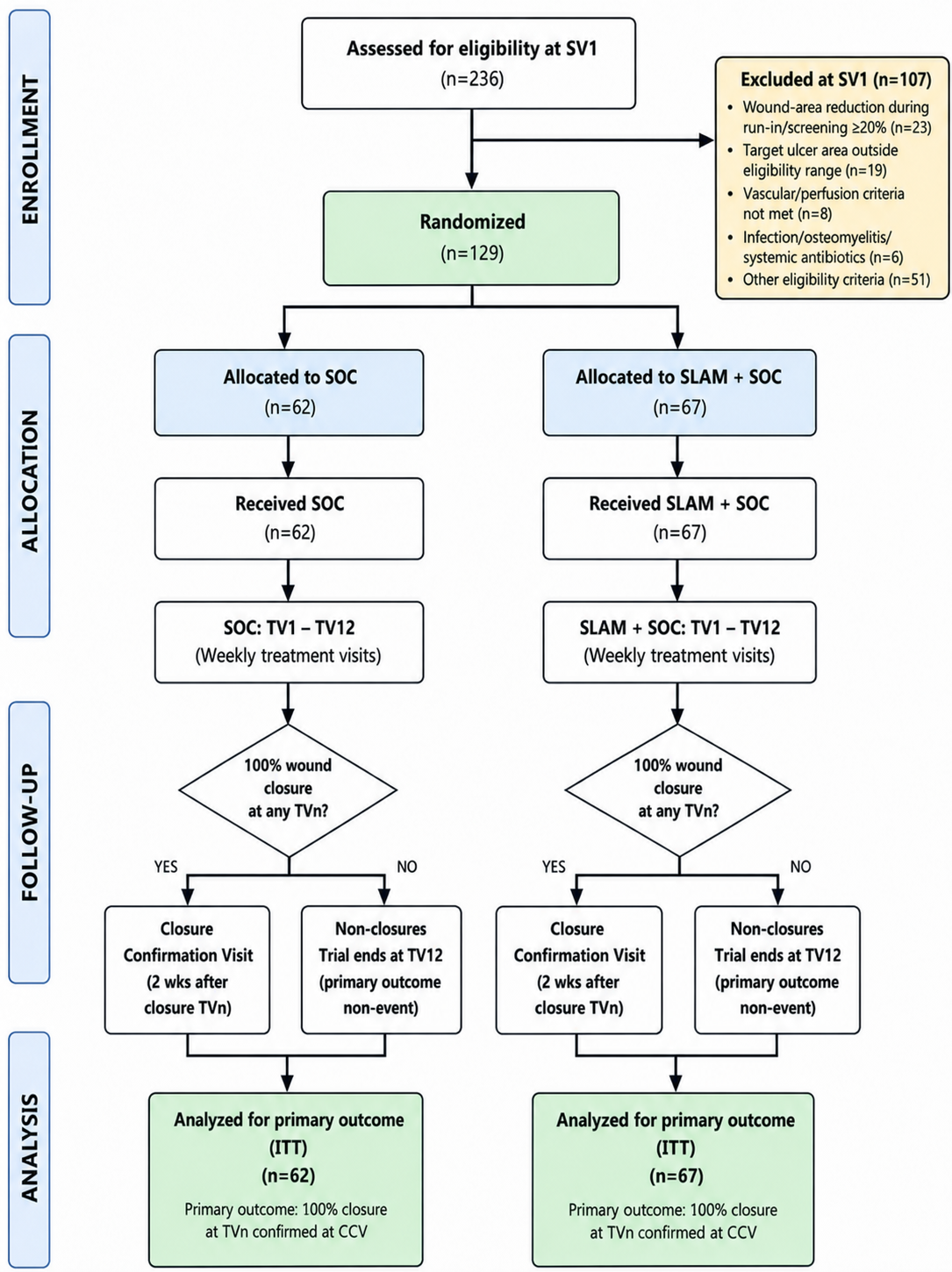
Figure 1. CAMPX CONSORT Diagram.
A total of 236 patients with DFUs from multiple sites were assessed for eligibility; 107 were excluded and 129 randomized. Overall, 62 patients received SOC alone, and 67 patients received SLAM plus SOC. At the interim data lock, 15 randomized participants remained ongoing and were considered ‘not healed’ in the ITT analysis; the remaining 114 had completed or otherwise exited follow-up by the data lock. The ITT closure analysis included all randomized participants (62 SOC; 67 SLAM plus SOC). The PP closure analysis included 60 participants who met protocol-required study product exposure criteria (28 SOC; 32 SLAM plus SOC). ITT PAR summaries included 120 participants after outlier exclusion (58 SOC; 62 SLAM plus SOC), and PP PAR summaries included 60 observations (28 SOC; 32 SLAM plus SOC). Summary statistics on demographic variables are provided in Table 3.
No statistically significant differences were observed across treatment groups (all p > 0.05) except race and Fitzpatrick Scale. Most measured baseline characteristics were similar between groups, although imbalances were observed for race and Fitzpatrick skin type. Wound area, wound age, and patient age were used as stratification factors in the trial design, and at this interim analysis, they are summarized descriptively to assess balance, shown in Table 4. The reported p-values are exploratory checks of randomization balance and were not used to adjust the interim analysis endpoints.
Table 3. Demographic Summary Statistics By Treatment Group.
| Variable | SOC | SLAM | P Value |
|---|---|---|---|
| Age (Years) | |||
|
61.8 ± 10.7 | 62.3 ± 12.9 | 0.677 |
| Gender, n/N (%) | |||
|
49/62 (79.0) | 51/67 (76.1) | 0.853 |
|
13/62 (21.0) | 16/67 (23.9) | - |
| Fitzpatrick Scale, n/N (%) | |||
|
12/62 (19.4) | 13/67 (19.4) | 0.02 |
|
27/62 (43.5) | 15/67 (22.4) | - |
|
16/62 (25.8) | 21/67 (31.3) | - |
|
4/62 (6.5) | 8/67 (11.9) | - |
|
3/62 (4.8) | 3/67 (4.5) | - |
|
0/62 (0.0) | 7/67 (10.4) | - |
| Wagner Grade, n/N (%) | |||
|
0/62 (0) | 0/67 (0) | 0.633 |
|
45/62 (72.6) | 45/67 (67.2) | - |
|
17/62 (27.4) | 22/67 (32.8) | - |
|
0/62 (0) | 0/67 (0) | - |
|
0/62 (0) | 0/67 (0) | - |
|
0/62 (0) | 0/67 (0) | - |
| Race, n/N (%) | |||
|
0/62 (0) | 0/67 (0) | 0.009 |
|
1/62 (1.6) | 0/67 (0) | - |
|
6/62 (9.7) | 21/67 (31.3) | - |
|
0/62 (0) | 0/67 (0) | - |
|
53/62 (85.5) | 46/67 (68.7) | - |
|
2/62 (3.2) | 0/67 (0) | - |
| Current Tobacco Use, n/N (%) | |||
|
5/62 (8.1) | 7/67 (10.4) | 0.871 |
|
57/62 (91.9) | 60/67 (89.6) | - |
SLAM–single layer amniotic membrane; SOC–standard of care
Table 4. Stratification Summary Statistics1.
| Variable | SOC | SLAM | P-Value |
|---|---|---|---|
| Wound area, n/N (%) | |||
|
24/59 (40.7) | 23/65 (35.4) | 0.717 |
|
35/59 (59.3) | 42/65 (64.6) | |
| Wound age, n/N (%) | |||
|
34/59 (57.6) | 35/65 (53.8) | 0.788 |
|
25/59 (42.4) | 30/65 (46.2) | |
| Patient age, n/N (%) | |||
|
26/59 (44.1) | 28/65 (43.1) | 0.857 |
|
33/59 (55.9) | 37/65 (56.8) | |
1For stratification summary statistics, 3 participants from SOC and 2 from SLAM did not provide stratification characteristic information. SLAM–single layer amniotic membrane; SOC–standard of care
The intention-to-treat (ITT) and per protocol (PP) populations were analyzed. The ITT population was defined as all randomized participants, analyzed according to their assigned treatment group. The PP population was defined as randomized participants who met the protocol-specified treatment-exposure requirements.
COMPLETE WOUND CLOSURE
The primary endpoint was assessed for the interim analysis. The primary endpoint was the percentage of target ulcers achieving complete wound closure in 12 weeks.
Complete wound closure was summarized using risk ratios with 95% confidence intervals. Due to the absence of a pre-specified interim alpha-spending plan, p-values are reported as unadjusted. In the ITT population, the SLAM plus SOC arm achieved a 17.9% (n=12/67) closure rate versus 11.3% (n=7/62) with SOC alone (RR 1.59, 95% CI: 0.67–3.77; two-sided Fisher’s exact p=0.329). Among the PP population, closure was 31.2% (n=10/32) on SLAM plus SOC versus 21.4% (n=6/28) on SOC (RR 1.46, 95% CI: 0.61–3.50; two-sided Fisher’s exact p=0.559).
PAR
Additionally, the percent area reduction (PAR) from TV-1 to TV-13 was measured with digital photographic planimetry using an imaging device, and wound measurements obtained by physical examination were analyzed weekly. PAR was calculated using wound surface area measurements obtained during post-debridement assessments. Baseline area was defined as the wound area measured at randomization, and subsequent PAR values were calculated relative to this baseline using measurements collected at each follow-up visit. Considering that wound area was measured after wound-bed preparation at each visit, PAR reflected changes in the clinically assessed wound bed over time under a standardized measurement procedure
Within each arm, any individual PAR value falling below Q1 - 1.5*IQR or above Q3 + 1.5*IQR was flagged and excluded. Each participant's final PAR value was used; for those that were still ongoing, their latest PAR was used. For ITT, SLAM plus SOC had numerically higher mean and median wound area reduction than SOC, with a mean PAR of 28.5% versus 24.3% (without outliers). Summary statistics are provided in Table 5.
Table 5. Percent Area Reduction (PAR) Summary Statistics without outliers for ITT.
| Treatment Arm | n | Mean |
Standard Deviation |
Median | IQR |
|---|---|---|---|---|---|
| SOC | 58 | 24.3 | 53.9 | 20.0 | 79.8 |
| SLAM plus SOC | 62 | 28.5 | 55.9 | 31.6 | 80.2 |
SLAM–single layer amniotic membrane; SOC–standard of care
For PP, SLAM plus SOC had higher mean and median wound area reductions SOC on both average and median wound area reduction, with summary statistics for each treatment group (without outliers) reported in Table 6.
Table 6. Percent Area Reduction (PAR) Summary Statistics without outliers for PP.
| Treatment Arm | n | Mean |
Standard Deviation |
Median | IQR |
|---|---|---|---|---|---|
| SOC | 28 | 38.0 | 60.3 | 55.1 | 103.8 |
| SLAM plus SOC | 32 | 47.4 | 63.7 | 69.2 | 79.2 |
SLAM–single layer amniotic membrane; SOC–standard of care
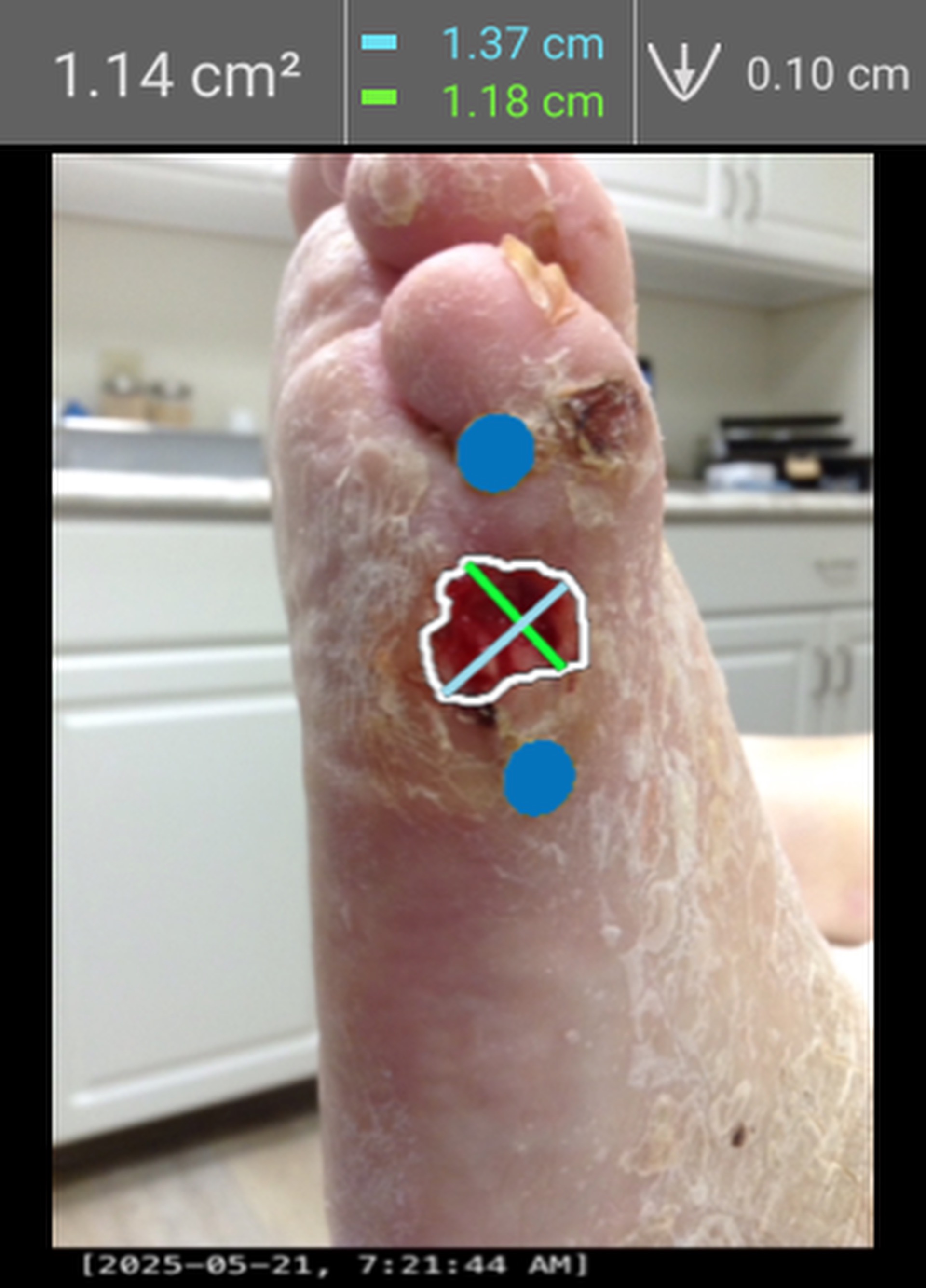
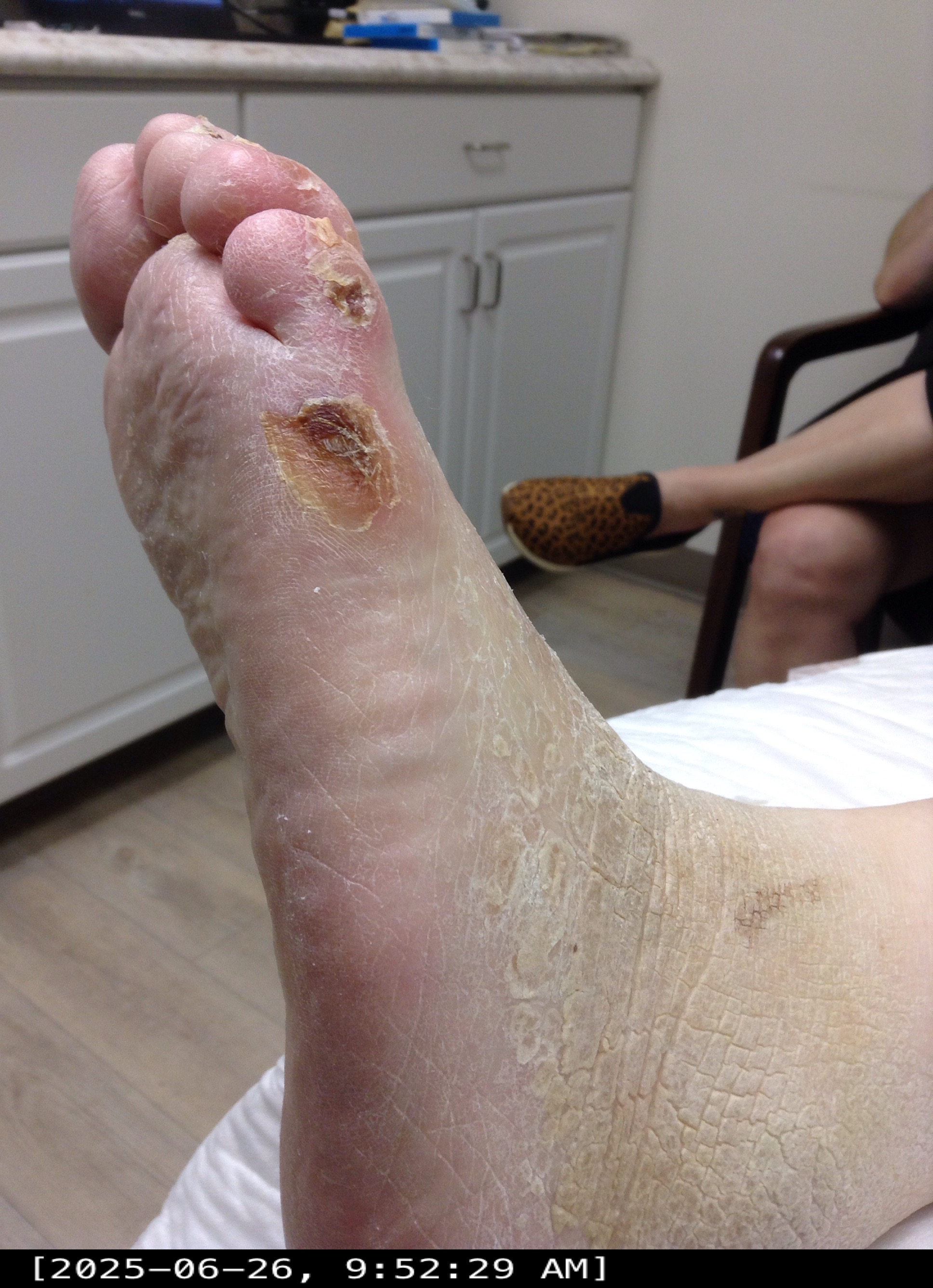
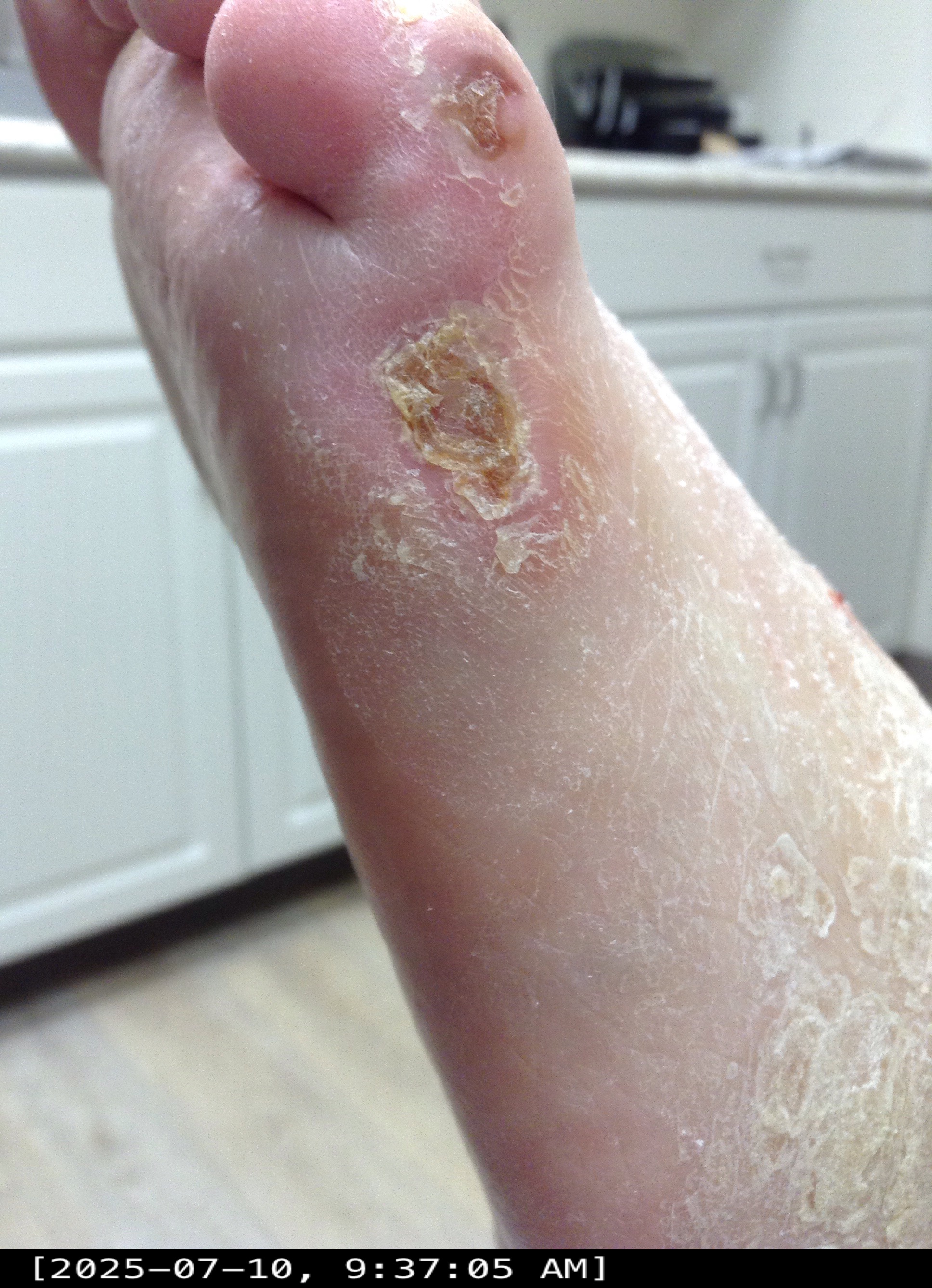
Sequential images shown in Figure 2 document the trajectory of wound healing from TV-1, TV-7, and CCV in a patient assigned to the SLAM plus SOC treatment arm.
 |
 |
 |
|---|
Figure 2. Digital images from TV-1, TV-7, and CCV (left to right), SLAM plus SOC treatment arm.
DISCUSSION
This planned interim analysis was conducted following a formal data lock of the electronic data capture (EDC) system and completion of a quality assurance review prior to statistical evaluation. The objectives of this interim assessment were to evaluate baseline comparability between treatment groups and to examine preliminary outcomes for the primary endpoint and PAR relative to SOC. Participants were stratified by baseline wound size, wound duration, and patient age. Although no substantial differences were observed across pre-specified stratification variables between treatment arms, imbalances in race and Fitzpatrick skin type were observed. Pre-specified subgroup analyses based on stratification factors will be performed as part of the final analysis.
For the primary endpoint, the SLAM plus SOC group demonstrated higher wound closure rates compared with SOC alone in both the ITT and PP populations. In the ITT population, the relative risk was 1.59 (95% CI: 0.67–3.77), while the PP population showed a relative risk of 1.46 (95% CI: 0.61–3.50). Enrollment is ongoing and will continue until the pre-specified sample size is reached for all treatment arms.
Analysis of PAR further informed treatment group comparisons. In both the ITT and PP populations, participants treated with SLAM in combination with SOC achieved a greater mean wound area reduction than those receiving SOC alone. These interim findings suggest favorable treatment trends and provide preliminary evidence of a numerical treatment trend, pending confirmation in the final analysis.
LIMITATIONS
Several limitations should be considered when interpreting these findings. The study was open-label with respect to treatment allocation, as blinding of participants and treating investigators was not feasible due to the nature of the interventions. At the time of this analysis, 15 participants remained ongoing. In addition, PAR outliers were excluded from the PAR analysis according to the prespecified analytic approach, which may influence estimates of wound area reduction. No adjustment for multiplicity was applied across secondary or exploratory analyses; therefore, these findings should be interpreted accordingly. Finally, incomplete or inconsistent follow-up among some participants may have affected the completeness of outcome ascertainment and introduced potential bias. Therefore, these results should be considered preliminary and hypothesis-generating, pending confirmation in the final analysis.
CONCLUSION
In this planned interim analysis of the CAMPX trial, adjunctive treatment with SLAM plus SOC produced numerically higher 12-week complete wound-closure rates than SOC alone in both the ITT population (17.9% versus 11.3%) and the PP population (31.2% versus 21.4%), in addition to greater mean and median percent area reduction. As a result of continued active enrollment, baseline imbalances, the treatment of participants still ongoing at the data lock as “not healed” in the ITT analysis, and outlier exclusions in the PAR analyses, these findings should be considered preliminary and should not be interpreted as establishing efficacy. Completion of enrollment and the pre-specified final analysis are needed to determine whether SLAM provides a reproducible benefit when added to SOC for chronic, non-healing DFUs.
Author Contributions: Conceptualization, T.S.; methodology, T.S., B.T., E.C., and D.S.; data curation, D.S.; writing–original draft preparation, T.S., B.T., E.C., and D.S.; writing–review and editing, T.S., B.T., E.C., and D.S.; visualization, T.S. and G.B.; project administration, R.B. All authors have read and agreed to the published version of the manuscript.
Received: 3 March 2026 | Accepted: 17 August 2026
Funding: This research was funded by Applied Biologics, LLC, including support for study operations, study materials, site coordination, data management infrastructure, and statistical analysis.
Institutional Review Board Statement: The protocol, informed consent form, and all subject-facing study materials were reviewed and approved by the appropriate Institutional Review Board (IRB)/Independent Ethics Committee (IEC) before study initiation. NCT06564831 is the ClinicalTrials.gov registration, and Pro00080075 is the IRB approval number.
Informed Consent Statement: Informed consent was obtained from all subjects involved in the study, including for the publication of wound images.
Data Availability Statement: De-identified participant-level data underlying the findings of this interim analysis may be made available by the corresponding author upon reasonable request, subject to sponsor approval, ongoing trial conduct considerations, and applicable data protection requirements.
Conflicts of Interest: This study was funded by Applied Biologics, LLC. The sponsor provided support for study operations, study materials, site coordination, data management infrastructure, and statistical analysis support. The sponsor participated in study design and operational oversight. SerenaGroup, Inc. received institutional funding from Applied Biologics, LLC, to conduct the study. T.S., B.T., E.C., D.S., G.B., and R.B. report no employment with, consulting arrangements with, honoraria from, stock or equity ownership in, or individual investigator payments from Applied Biologics, LLC. The authors report no other financial relationships with Applied Biologics, LLC or related wound care companies relevant to this work. Investigators retained responsibility for clinical conduct, interpretation of findings, manuscript preparation, and publication decisions. All authors had access to the interim dataset included in this report and accept responsibility for the integrity and accuracy of the analyses presented.
Declaration of Generative AI: During the preparation of this work the authors used OpenAI to support drafting and language refinement of portions of this manuscript, including improving clarity, grammar, and flow. The AI tool was not used for data generation, data analysis, interpretation of results, or drawing scientific conclusions. After using this tool/service, the authors reviewed and edited the content as needed and take full responsibility for the content of the publication.
Disclaimer/Publisher’s Note: The statements, opinions and data contained in all publications are solely those of the individual author(s) and contributor(s) and not of CWCMR and/or the editor(s). CWCMR and/or the editor(s) disclaim responsibility for any injury to people or property resulting from any ideas, methods, instructions or products referred to in the content.
REFERENCES
-
McDermott K , Fang M, Boulton AJM, Selvin E, Hicks CW. Etiology, Epidemiology, and Disparities in the Burden of Diabetic Foot Ulcers. Diabetes Care. 2023; 46(1): 209–221. https://doi.org/10.2337/dci22-0043
-
Dawi J, Tumanyan K, Tomas K, Misakyan Y, et al. Diabetic Foot Ulcers: Pathophysiology, Immune Dysregulation, and Emerging Therapeutic Strategies. Biomedicines. 2025; 13(1076). https://doi.org/10.3390/biomedicines13051076
-
Game F. Classification of diabetic foot ulcers. Diabetes Metab Res Rev. 2016; 32(1): 186–94. https://doi.org/10.1002/dmrr.2746
-
Ben Zvi L, Maman D, Margulis M, Berkovich Y. Predictors of Major Amputation and Mortality in Infected Diabetic Foot Ulcers: A Retrospective Nationwide Inpatient Sample Study. Int. J. Environ. Res. Public Health. 2025; 22(9):1387. https://doi.org/10.3390/ijerph22091387
-
Fife CE, Eckert KA, Carter MJ. Publicly Reported Wound Healing Rates: The Fantasy and the Reality. Adv Wound Care. 2018; 7(3):77–94. https://doi.org/10.1089/wound.2017.0743
-
Lim JZM, Ng NSL, Thomas C. Prevention and treatment of diabetic foot ulcers. J R Soc Med. 2017; 110(3): 104–109. https://doi.org/10.1177/0141076816688346
-
Bradford Rice J, Desai U, Cummings AKG, Birnbaum HG, et al. Burden of Diabetic Foot Ulcers for Medicare and Private Insurers. Diabetes Care 2014; 37(3): 651–658. https://doi.org/10.2337/dc13-2176
-
Wu S, Carter M, Cole W, Crombie R, et al. Best practice for wound repair and regeneration use of cellular, acellular and matrix-like products (CAMPs). J Wound Care. 2023; 32(Sup4b): S1–S31. https://doi.org/10.12968/jowc.2023.32.Sup4b.S1
-
Mohammed YA, Farouk HK, Gbreel MI, et al. Human amniotic membrane products for patients with diabetic foot ulcers: do they help? A systematic review and meta-analysis. J Foot Ankle Res. 2022; (15):71. https://doi.org/10.1186/s13047-022-00575-y
-
Laurent I, Astère M, Wang KR, et al. Efficacy and Time Sensitivity of Amniotic Membrane treatment in Patients with Diabetic Foot Ulcers: A Systematic Review and Meta-analysis. Diabetes Ther. 2017 ;8(5):967–79. https://doi.org/10.1007/s13300-017-0298-8